. 2005 May;115(5):1188-98.
doi: 10.1172/JCI23371. Epub 2005 Apr 1.

Synaptopodin regulates the actin-bundling activity of alpha-actinin in an isoform-specific manner

Affiliations

Synaptopodin regulates the actin-bundling activity of alpha-actinin in an isoform-specific manner

Katsuhiko Asanuma et al. J Clin Invest. 2005 May.

Erratum in

  • J Clin Invest. 2012 Feb 1;122(2):781

Abstract

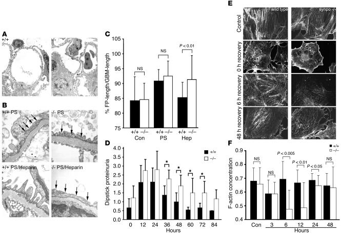
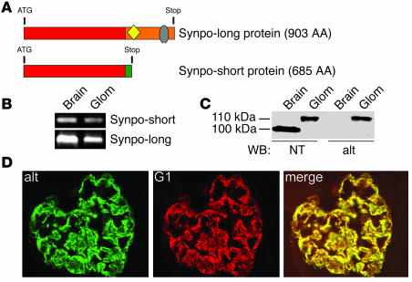
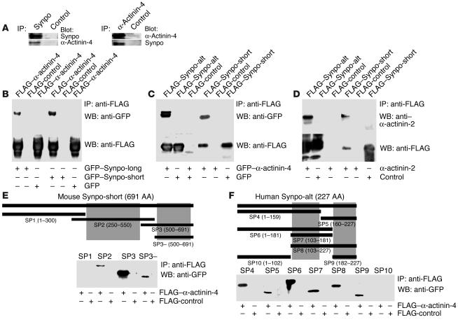
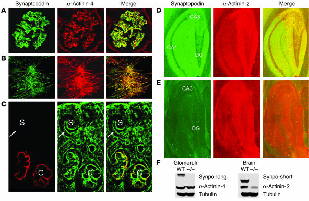
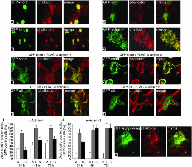
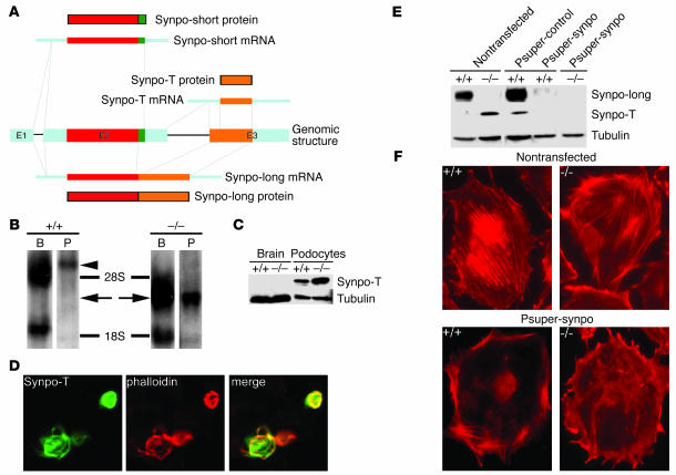
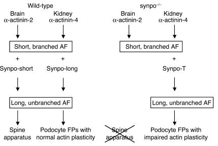
Synaptopodin is the founding member of a novel class of proline-rich actin-associated proteins highly expressed in telencephalic dendrites and renal podocytes. Synaptopodin-deficient (synpo(-/-)) mice lack the dendritic spine apparatus and display impaired activity-dependent long-term synaptic plasticity. In contrast, the ultrastructure of podocytes in synpo(-/-) mice is normal. Here we show that synpo(-/-) mice display impaired recovery from protamine sulfate-induced podocyte foot process (FP) effacement and LPS-induced nephrotic syndrome. Similarly, synpo(-/-) podocytes show impaired actin filament reformation in vitro. We further demonstrate that synaptopodin exists in 3 isoforms, neuronal Synpo-short (685 AA), renal Synpo-long (903 AA), and Synpo-T (181 AA). The C terminus of Synpo-long is identical to that of Synpo-T. All 3 isoforms specifically interact with alpha-actinin and elongate alpha-actinin-induced actin filaments. synpo(-/-) mice lack Synpo-short and Synpo-long expression but show an upregulation of Synpo-T protein expression in podocytes, though not in the brain. Gene silencing of Synpo-T abrogates stress-fiber formation in synpo(-/-) podocytes, demonstrating that Synpo-T serves as a backup for Synpo-long in synpo(-/-) podocytes. In concert, synaptopodin regulates the actin-bundling activity of alpha-actinin in highly dynamic cell compartments, such as podocyte FPs and the dendritic spine apparatus.


Figures

Figure 1
Figure 1
Delayed actin filament reformation in synpo–/– podocytes. (A) By TEM analysis, podocytes of synpo–/– mice appear structurally normal (magnification, ×20,000). +/+, wild-type; –/–, synpo–/–. (B) Impaired recovery of synpo–/– mice from PS-induced FP effacement. Top: comparable degrees of effacement (arrows) in wild-type and synpo–/– mice. Bottom: complete recovery after heparin treatment in wild-type but not synpo–/– mice (magnification, ×40,000). (C) Quantitative analysis showing significantly delayed FP re-formation in synpo–/– mice during recovery induced by heparin sulfate (Hep) (P < 0.01). Con, control. (D) LPS injection causes delayed recovery from proteinuria in synpo–/– mice. Statistically significant changes were found between 36 and 72 hours; *P < 0.05. (E) Confocal imaging showing delayed re-formation of stress fibers in synpo–/– podocytes (magnification, ×600). No changes were found between wild-type and synpo–/– podocytes before and directly after actin depolymerization with cytochalasin D (0 hours recovery). After washout of cytochalasin D, actin fibers were fully reestablished in wild-type cells after 6 hours. In synpo–/– podocytes, stress fiber formation did not return to control levels before 48 hours. (F) Quantitative analysis of F-actin content in wild-type and synpo–/– podocytes. No significant differences were found in untreated controls and at 3 hours after removal of cytochalasin D. Wild-type cells returned to control levels at 6 hours. In contrast, synpo–/– cells displayed significantly reduced F-actin content at 6, 12, and 24 hours after washout. At 48 hours, synpo–/– mice displayed wild-type level.
Figure 2
Figure 2
Synaptopodin isoform expression in kidney and brain. (A) Comparison of Synpo-long and Synpo-short. The red box shows the first 670 AA that are shared between both isoforms; the orange box shows AA 671–903 corresponding to the Synpo-alt fragment of Synpo-long. Synpo-alt contains a LPPPP motif (yellow) for ENA/VASP binding and a PPRPF motif (gray) for homer binding. (B) RT-PCR with isoform-specific primers reveals the expression of both Synpo-short and Synpo-long mRNA in brain and kidney glomeruli (Glom). (C) Western blotting (WB) with the polyclonal NT antibody against an epitope present in both Synpo-long and Synpo-short and the Synpo-alt–specific antibody (alt). At the protein level, only Synpo-long is expressed in the kidney, whereas only Synpo-short is expressed in the brain. (D) Double-labeling immunofluorescence microcopy of Synpo-alt with the pan-synaptopodin monoclonal antibody G1 confirmed the expression of Synpo-long in podocytes (magnification, ×650).
Figure 3
Figure 3
Synpo-short and Synpo-long interact with α-actinin-4 and α-actinin-2. (A) Coimmunoprecipitation experiments showing that endogenous synaptopodin interacts with α-actinin-4 in podocytes. Left: Immunoprecipitation with anti-synaptopodin (Synpo). Right: Immunoprecipitation with anti–α-actinin-4. No interaction was found with a control antibody. (B) GFP-tagged Synpo-long and Synpo-short coimmmunoprecipitated with FLAG-tagged α-actinin-4 but not with the FLAG control. (C) In a converse experiment, GFP-tagged α-actinin-4 coprecipitated with FLAG–Synpo-alt and FLAG–Synpo-short. No interaction was found with the GFP control. (D) FLAG–Synpo-alt and FLAG–Synpo-short also interact with GFP-tagged α-actinin-2. (E) Synpo-short contains 2 independent nonoverlapping interaction sites (gray boxes) for α-actinin-4 that are contained in AA 300–550 and 550–691. In contrast, the SP1 fragment and the FLAG control do not interact with α-actinin. (F) Synpo-alt also contains 2 independent nonoverlapping α-actinin interaction sites that are localized in AA 103–159 and 182–227 of the Synpo-alt fragment. Hence, Synpo-long contains 4 α-actinin interacting sites, whereas Synpo-short contains only 2 α-actinin interacting sites.
Figure 4
Figure 4
Synaptopodin colocalizes with α-actinin in tissues and cultured cells. (A) In the adult kidney, synaptopodin colocalizes with α-actinin-4 in podocytes (magnification, ×650). (B) Synaptopodin colocalizes with α-actinin-4 along actin filaments in differentiated cultured wild-type podocytes (magnification, ×650). (C) During kidney development, α-actinin-4 is already found in undifferentiated podocytes (arrows) of the S-shaped body stage (S) that have a cortical actin cytoskeleton. In contrast, synaptopodin is first found in the capillary loop stage (C) when podocytes start to develop long, unbranched contractile actin bundles (magnification, ×450). (D) Codistribution of synaptopodin with α-actinin-2 in the CA1, CA3, and dendate gyrus (DG) regions in the hippocampus of a 6-month-old wild-type mouse (magnification, ×650). (E) Lack of synaptopodin and downregulation of α-actinin-2 labeling in all regions of the hippocampus in an age-matched synpo–/– mouse (magnification, ×650). (F) Western blot analysis of cytosolic extract from glomeruli (left) and brains (right) of 6-month-old wild-type and synpo–/– mice. The levels of α-actinin-4 are not different between wild-type and synpo–/– mice. In contrast, there is a strong downregulation of α-actinin-2 in the brains of synpo–/– mice. Equal protein loading was confirmed by reprobing for tubulin.
Figure 5
Figure 5
Synaptopodin cooperates with α-actinin in the elongation and bundling of α-actinin–induced actin filaments. (A) GFP–Synpo-short (GFP-short) or (B) GFP–Synpo-long (GFP-long) induce and colocalize with amorphous cytoplasmic phalloidin-positive aggregates, suggesting the presence of an actin binding site. (C) GFP–Synpo-alt (GFP-alt) does not induce phalloidin-positive aggregates, showing that the actin binding site is localized in the first 670 AA that are shared between Synpo-long and Synpo-short. (D) Single transfection of α-actinin-4 induces short, branched actin filaments. (E) Cotransfection of GFP–Synpo-short with FLAG–α-actinin-4 converts the short, branched filaments into long, parallel, unbranched actin bundles. (F) Inhibition of actin-branching activity of FLAG–α-actinin-4 is also observed after cotransfection with GFP–Synpo-alt. (G) Cotransfection of GFP–Synpo-short with FLAG–α-actinin-2 also induces long, unbranched actin bundles. (H) In contrast, cotransfection of GFP–Synpo-alt with FLAG–α-actinin-2 does not affect α-actinin-2–induced short, branched cortical actin filaments. (I and J) Quantitative analysis of actin-bundling kinetics. The data are presented as percentage of actin bundle–positive cells per GFP-positive cells. (I) Cells coexpressing α-actinin-4 and GFP–Synpo-long (L) show faster kinetics of actin bundling at 24, 48, and 72 hours than cells expressing GPF–Synpo-alt (A) or GFP–Synpo-short (S). (J). After cotransfection with α-actinin-2, GFP–Synpo-long and GFP–Synpo-short show comparable kinetics, whereas virtually no actin-bundling activity is detected at any time points in cells coexpressing α-actinin-2 and GFP–Synpo-alt. (K) Transfection of undifferentiated wild-type podocytes with GFP–Synpo-long induces stress fibers similar to those found in differentiated wild-type cells (see Figure 4B). No stress fibers are found in nontransfected cells (asterisk). Magnification, ×600 (AH); ×450 (K).
Figure 6
Figure 6
Synpo-T serves as a backup in podocytes but not in brains of synpo–/– mice. (A) Genomic organization and alternative splice products of mouse synaptopodin. In addition to Synpo-long and Synpo-short, a third isoform, Synpo-T, is generated from exon 3. Synpo-T encodes a protein that is identical to AA 721–901 of Synpo-long. E1–E3, exons 1–3. (B) Northern blot analysis showing the induction of Synpo-T mRNA (arrows) in brains (B) and cultured podocytes (P) of synpo–/– mice. In wild-type mice, the Synpo-T probe cross-hybridizes with Synpo-long (arrowhead). (C) Western blot analysis showing the upregulation of Synpo-T protein expression in synpo–/– podocytes. In contrast, Synpo-T is not expressed at the protein level in the brain. (D) Synpo-T is sufficient to convert FLAG–α-actinin-4–induced short, branched filaments into long, parallel, unbranched actin bundles (magnification, ×450). (E) Western blot analysis of Synpo-long and Synpo-T before (nontransfected) and after gene silencing by stable expression of Synpo-long– and Synpo-T–specific siRNA (Psuper-synpo). Psuper-synpo but not a control siRNA construct (Psuper-control) abrogates Synpo-long and Synpo-T protein expression in wild-type and synpo–/– podocytes. Equal protein loading was assessed by reprobing for tubulin. (F) Phalloidin staining reveals well-developed stress fibers in nontransfected wild-type and synpo–/– podocytes. In contrast, wild-type and synpo–/– podocytes expressing Psuper-synpo are devoid of stress fibers (magnification, ×650).
Figure 7
Figure 7
Working model of the cellular function of synaptopodin. In the wild-type brain, Synpo-short converts α-actinin-2–induced short, branched actin filaments (AF) into long, unbranched actin filaments that are found between the ER stacks of the spine apparatus. Similarly, Synpo-long converts short, cortical α-actinin–induced actin filaments of immature podocytes into long, unbranched contractile filaments found in FPs of differentiated podocytes. In the brains of synpo–/– mice, Synpo-T is not expressed, leading to the loss of the spine apparatus. In contrast, Synpo-T is upregulated in synpo–/– podocytes and can partially rescue the loss of Synpo-long in podocytes, albeit at the price of a delayed plasticity of the actin filaments.

Similar articles

Cited by

References

    1. Mundel P, Gilbert P, Kriz W. Podocytes in glomerulus of rat kidney express a characteristic 44 KD protein. J. Histochem. Cytochem. 1991;39:1047–1056. - PubMed
    1. Mundel P, et al. Synaptopodin: an actin-associated protein in telencephalic dendrites and renal podocytes. J. Cell Biol. 1997;139:193–204. - PMC - PubMed
    1. Weins A, et al. Differentiation- and stress-dependent nuclear cytoplasmic redistribution of myopodin, a novel actin-bundling protein. J. Cell Biol. 2001;155:393–404. - PMC - PubMed
    1. Deller T, Merten T, Roth SU, Mundel P, Frotscher M. Actin-associated protein synaptopodin in the rat hippocampal formation: localization in the spine neck and close association with the spine apparatus of principal neurons. J. Comp. Neurol. 2000;418:164–181. - PubMed
    1. Deller T, et al. Synaptopodin-deficient mice lack a spine apparatus and show deficits in synaptic plasticity. Proc. Natl. Acad. Sci. U. S. A. 2003;100:10494–10499. - PMC - PubMed

Publication types