. 2005 May;115(5):1267-74.
doi: 10.1172/JCI22782. Epub 2005 Apr 14.

The N-terminal domain of thrombomodulin sequesters high-mobility group-B1 protein, a novel antiinflammatory mechanism

Affiliations

The N-terminal domain of thrombomodulin sequesters high-mobility group-B1 protein, a novel antiinflammatory mechanism

Kazuhiro Abeyama et al. J Clin Invest. 2005 May.

Abstract

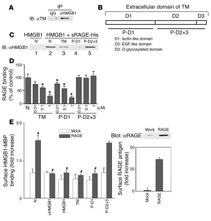
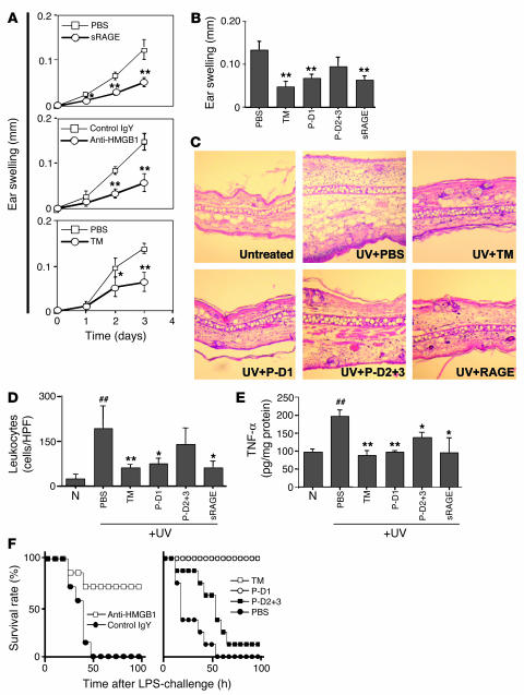
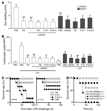
Thrombomodulin (TM) is an endothelial anticoagulant cofactor that promotes thrombin-mediated formation of activated protein C (APC). We have found that the N-terminal lectin-like domain (D1) of TM has unique antiinflammatory properties. TM, via D1, binds high-mobility group-B1 DNA-binding protein (HMGB1), a factor closely associated with necrotic cell damage following its release from the nucleus, thereby preventing in vitro leukocyte activation, in vivo UV irradiation-induced cutaneous inflammation, and in vivo lipopolysaccharide-induced lethality. Our data also demonstrate antiinflammatory properties of a peptide spanning D1 of TM and suggest its therapeutic potential. These findings highlight a novel mechanism, i.e., sequestration of mediators, through which an endothelial cofactor, TM, suppresses inflammation quite distinctly from its anticoagulant cofactor activity, thereby preventing the interaction of these mediators with cell surface receptors on effector cells in the vasculature.


Figures

Figure 1
Figure 1
TM, via D1, binds to HMGB1. (A) For IP, rhs-TM (10 nM) and HMGB1 (10 nM) were mixed and incubated, followed by the addition of protein A agarose beads conjugated to anti-HMGB1 IgG (αHMGB1) or nonimmune IgG (IgG). Immunoprecipitates were solubilized, and reduced SDS-PAGE (10%) was followed by immunoblotting with anti-TM antibody (αTM). (B) TM extracellular domains. (C) Competition between TM and sRAGE for binding to HMGB1. First, sRAGE-His (10 nM) was incubated with HMGB1 alone (10 nM; lane 2) or in the presence of rhs-TM (TM, 1 μM; lane 3), P-D1 (1 μM; lane 4), or P-D2+3 (1 μM; lane 5). Then, nickel resin beads were added, precipitates were solubilized, and SDS-PAGE (10%) was followed by IB with αHMGB1 IgG. N, untreated controls. (D) Binding of sRAGE-His (10 nM) to HMGB1 immobilized on plastic plates was studied with untreated controls, TM, P-D1, or P-D2+3. (E) Left panel: Binding of HMGB1-MBP (1 nM) to RAGE-transfected (RAGE) or mock-transfected (Mock) COS-7 cells (104 cells/well) was assessed in the presence of untreated controls, αHMGB1, HMGB1, TM, P-D1, or P-D2+3 (100 nM each). Results shown are representative of 4 replicate wells. The right panel shows RAGE expression in RAGE-transfected versus mock-transfected COS-7 cells by IB (upper) and immunofluorescence on nonpermeabilized fixed cells (lower). *P < 0.05, compared with the control group. #P < 0.05, compared with the paired, untreated controls.
Figure 2
Figure 2
TM, via D1, prevents proinflammatory effects of HMGB1 in vitro. (A) Effect of HMGB1 on NF-κB–dependent gene transcription. THP-1 cells were transfected with an NF-κB promoter-reporter (SEAP) construct. Then, THP-1 cells (106 cells/ml) were incubated for 16 hours at 37°C with the indicated concentration of HMGB1, and reporter expression was determined. Recombinant sRAGE (10 nM) or rhs-TM (TM; 100 nM) was added as indicated. (B) Human peripheral blood mononuclear phagocytes (106 cells/ml) were loaded with DCF-DA and were incubated for 1 hour at 37°C in medium alone (N) or with HMGB1 (10 nM) in medium. As indicated, rhs-TM, P-D1, P-D2+3, E456, or sRAGE was added (100 nM each). Then, the formation of DCF was determined. *P < 0.05 and **P < 0.01, compared with untreated controls. #P < 0.05, compared with the paired control group without sRAGE treatment.
Figure 3
Figure 3
Central role of HMGB1 in UV irradiation–induced skin injury and the effect of TM and TM-derived peptides on HMGB1-mediated local and systemic inflammation. (A) In vivo effect of HMGB1 blockade on UV irradiation–induced cutaneous inflammation. The time course for ear swelling was measured after local subcutaneous administration of PBS or sRAGE (3 pmol; top); anti-HMGB1 IgY (Anti-HMGB1) or nonimmune IgY (Control IgY, 300 ng; middle); or rhs-TM (3 pmol) or PBS before exposure to UV irradiation (bottom). In each case, data shown are mean ± SD (n = 8). *P < 0.05; **P < 0.01. (BE) As indicated, mice received PBS, rhs-TM, P-D1, P-D2+3 (each at 100 nmol/kg; i.p.) or sRAGE (25 nmol/kg; i.p.) at 1 hour and 12 hours after exposure to UV irradiation (n = 8 per group). Three days after UV treatment, ear swelling (B), general histopathological features of the skin (C; H&E staining; magnification, ×100), infiltrating leukocytes (D; cells/HPF), and local TNF-α accumulation (E) were assessed. The mean ± SD of 6 determinations is shown (B, D, and E). *P < 0.05 and **P < 0.01, compared with the UV-irradiated control group given PBS. ##P < 0.01, compared with untreated controls. (F) Effect of TM and TM-derived peptides on systemic LPS-induced lethal challenge. Left panel: mice (BALB/c) received LPS (10 mg/kg; i.p.) in the presence of anti-HMGB1 IgY or nonimmune IgY (3 i.p. doses of 2 mg/kg/mouse of IgY at 2, 12, and 24 hours after LPS). Right panel: mice were treated with LPS as described above and also received rhs-TM, P-D1, P-D2+3, or PBS (3 i.p. doses of 100 nmol/kg/mouse for each peptide).
Figure 4
Figure 4
RAGE-dependent and -independent mechanism of HMGB1-mediated events and the inhibitory effects of TM and TM-derived peptides. (A and B) Skin inflammation was induced in the mouse ear using UV irradiation. C57/BL6 mice (RAGE+/+; white bars) received rhs-TM alone, sRAGE alone, or TM-derived peptides (each at 100 nmol/kg; i.p.) combined with sRAGE (25 nmol/kg; i.p.) at 1 hour and 12 hours after exposure to UV irradiation (n = 8 per group). RAGE-null mice (C57BL/6 strain; black bars) were UV irradiated and were treated with sRAGE (25 nmol/kg), rhs-TM, or TM-derived peptides (each at 100 nmol/kg; i.p.). After 3 days, ear swelling (A) and infiltrating leukocytes (B) were assessed. *P < 0.05 and **P < 0.01, compared with UV-irradiated, PBS-treated controls. #P < 0.05 and ##P < 0.01, compared with UV-irradiated RAGE-null mice. (C and D) Mice (wild-type C57BL/6 and RAGE-null on the C57BL/6 background) received LPS (10 mg/kg; i.p.) alone or in the presence of TM, P-D1, P-D2+3, and PBS. (C) Effect of rhs-TM or TM-derived peptides on LPS-induced lethality. Mice received 3 i.p. doses (100 nmol/kg/mouse) of anti-HMGB1 or control IgY at 2, 12, and 24 hours after LPS challenge. (D) Mice were treated with LPS and also received either anti-HMGB1 IgY or nonimmune IgY (3 i.p. doses of 2 mg/kg/mouse of IgY at 2, 12, and 24 hours after LPS). In C and D, (–/–) indicates that RAGE-null mice were used, and (+/+) indicates that strain-matched controls were used.

References

    1. Esmon CT, Owen GW. Identification of an endothelial cell cofactor for thrombin-catalyzed activation of protein C. Proc. Natl. Acad. Sci. U. S. A. 1981;78:249–252. - PMC - PubMed
    1. Esmon CT, Esmon NL, Harris KW. Complex formation between thrombin and thrombomodulin inhibits both thrombin-catalyzed fibrin formation and factor V activation. J. Biol. Chem. 1982;257:7944–7947. - PubMed
    1. Maruyama I, Salem HH, Majerus PW. Coagulation factor Va binds to human umbilical vein endothelial cells and accelerates protein C activation. J. Clin. Invest. 1984;74:224–230. - PMC - PubMed
    1. Esmon CT. The roles of protein C and thrombomodulin in the regulation of blood coagulation. J. Biol. Chem. 1989;264:4743–4746. - PubMed
    1. Suzuki K, et al. Structure and expression of human thrombomodulin, a thrombin receptor on endothelium acting as a cofactor for protein C activation. EMBO J. 1987;6:1981–1997. - PMC - PubMed

Publication types