. 1992 Jun 11;326(24):1599-607.
doi: 10.1056/NEJM199206113262404.

The frequency of uniparental disomy in Prader-Willi syndrome. Implications for molecular diagnosis

Affiliations

The frequency of uniparental disomy in Prader-Willi syndrome. Implications for molecular diagnosis

M J Mascari et al. N Engl J Med. .

Abstract

Background: Prader-Willi syndrome is a genetic disorder characterized by infantile hypotonia, obesity, hypogonadism, and mental retardation, but it is difficult to diagnose clinically in infants and young children. In about two thirds of patients, a cytogenetically visible deletion can be detected in the paternally derived chromosome 15 (15q11q13). Recently, patients with Prader-Willi syndrome have been described who do not have the cytogenetic deletion but instead have two copies of the 15q11q13 region that are inherited from the mother (with none inherited from the father). This unusual form of inheritance is known as maternal uniparental disomy. Using molecular genetic techniques, we sought to determine the frequency of uniparental disomy in Prader-Willi syndrome.

Methods: We performed molecular analyses using DNA markers within 15q11q13 and elsewhere on chromosome 15 in 30 patients with Prader-Willi syndrome who had no cytogenetically visible deletion. We also studied their parents. Three patients with Prader-Willi syndrome who had a cytogenetic deletion served as controls.

Results: In 18 of the 30 patients without a cytogenetic deletion (60 percent), we demonstrated the presence of maternal uniparental disomy for chromosome 15 and its association with advanced maternal age. In another eight patients (27 percent), we identified large molecular deletions. The remaining four patients (13 percent) had evidence of normal biparental inheritance for chromosome 15; three of these patients were the only ones in the study who had some atypical clinical features.

Conclusions: In about 20 percent of all cases, Prader-Willi syndrome results from the inheritance of both copies of chromosome 15 from the mother (maternal uniparental disomy). With the combined use of cytogenetic and molecular techniques, the genetic basis of Prader-Willi syndrome can be identified in up to 95 percent of patients.


Figures
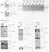
Figure 1.
Figure 1.
Representative Autoradiographs Obtained by Molecular Analysis of DNA Patients with Prader-Willi Syndrome with DNA Probes Specific for Chromosome 15q11q13. With the exception of Panel B, the various enzymes used are given in parentheses; HindIII was used with all probes shown in Panel B. The sizes of the DNA fragments are listed in kilobases (kb). C represents a constant, nonpolymorphic fragment. Probands are denoted by solid symbols. Panel A shows paternal deletions by RFLPs in two families. Both probands (lane 1) receive a single copy of a maternal allele (lane 3), but no paternal alleles (lane 2). In Panel B, the number of copies per genome of chromosome 15q11q13-specific probes is compared with the number of copies per genome of a chromosome 13 control probe (H2-26). For each proband, a deletion is indicated by a minus sign, and a two-copy hybridization by a plus sign. Panel C shows uniparental disomy in the monozygotic twins, as detected by probe 189-1 (upper autoradiograph). Use of probe IR10-1 (lower autoradiograph) showed that the specific type of uniparental disomy present was isodisomy, whereas probe 34 was uninformative. Panel D shows uniparental disomy in two additional probands with Prader–Willi syndrome, as detected by probe IR10-1, and heterozygosity (parental origin undetermined), as detected by probes 189-1 and 34.
Figure 2.
Figure 2.
Patterns of Uniparental Disomy in 18 Patients with Prader–Willi Syndrome. The order of seven DNA probes within chromosome 15q11q13 and three DNA probes distal to this region is shown from the centromere (cen) to the telomere (tel),,, (and unpublished data). Brackets indicate that the order of probes is unknown. The loci defined are listed above the probes. Four alternative molecular classes of deletions in Prader–Willi syndrome, as discussed in the text, are shown (deletion region). DNA probes MS1-14 and DP151 map to 15q14–q22, whereas MS620 maps close to the telomere. Cytogenetic heteromorphisms (15p/cen) are noted. Patient 1 was not studied with probe MS620, and locus IR39d was not evaluated in Patients 3, 4, 5, 6, and 29. In addition, not all families were evaluated with probes MS1-14 and DP151. Patient 24 demonstrated maternal uniparental disomy after digestion of DNA with KpnI and was found to be heterozygous with the use of StyI at the IR4-3R locus, and Patient 3 was found to be heterozygous with the use of PvuII at the IR4-3R locus (unpublished data).
Figure 3.
Figure 3.
Detection of Maternal Uniparental Disomy in Prader–Willi Syndrome with a Highly Informative Chromosome 15 DNA Probe. Autoradiographs are shown for probe MS620, on AluI-digested DNA from representative families with a proband with Prader–Willi syndrome. The sizes of DNA standards are listed on the right.

References

    1. Cassidy SB. Prader-Willi syndrome. Curr Probl Pediatr 1984;14:1–55. - PubMed
    1. Butler MG. Prader-Willi syndrome: current understanding of cause and diagnosis. Am J Med Genet 1990;35:319–32. - PMC - PubMed
    1. Greenberg F, Elder FFB, Ledbetter DH. Neonatal diagnosis of Prader-Willi syndrome and its implications. Am J Med Genet 1987;28:845–56. - PubMed
    1. Aughton DJ, Cassidy SB. Physical features of Prader-Willi syndrome in neonates. Am J Dis Child 1990;144:1251–4. - PubMed
    1. Hawkey CJ, Smithies A. The Prader-Willi syndrome with a 15/15 translocation: case report and review of the literature. J Med Genet 1976; 13:152–7. - PMC - PubMed

Publication types