Comparative Study
. 2005 May;132(10):2377-88.
doi: 10.1242/dev.01834. Epub 2005 Apr 20.

The retinoblastoma gene pathway regulates the postmitotic state of hair cells of the mouse inner ear

Affiliations
Comparative Study

The retinoblastoma gene pathway regulates the postmitotic state of hair cells of the mouse inner ear

Johanna Mantela et al. Development. 2005 May.

Abstract

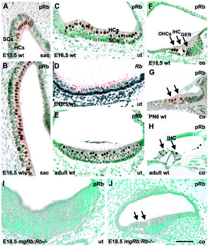
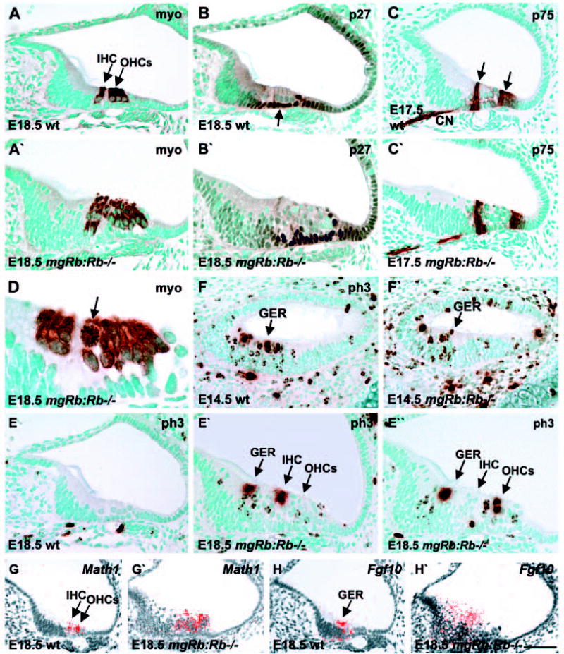
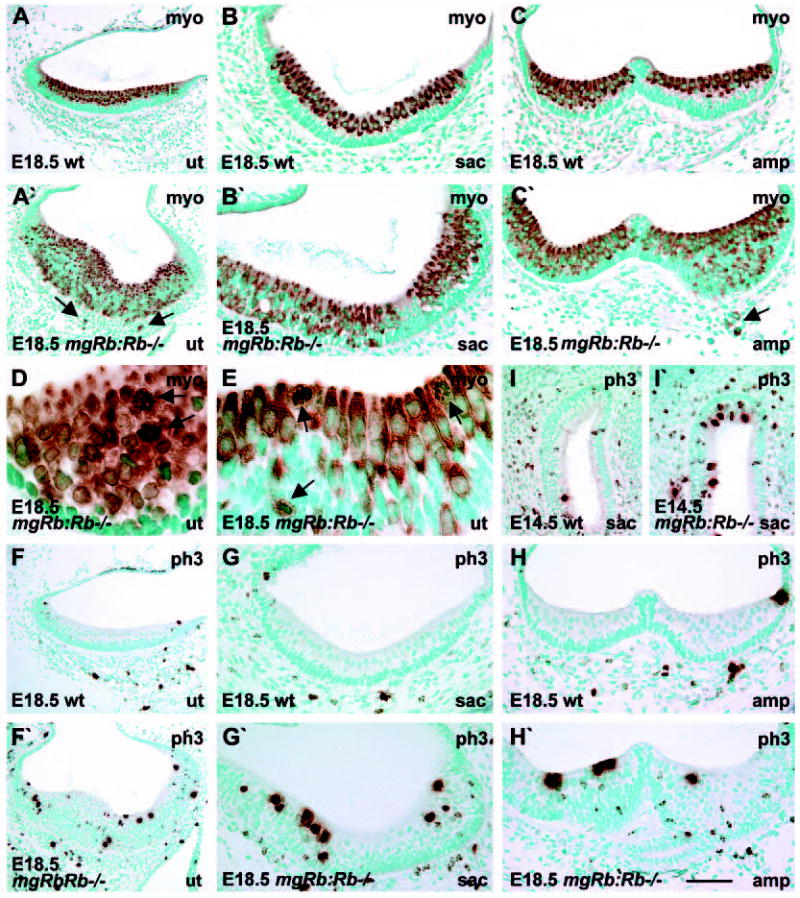
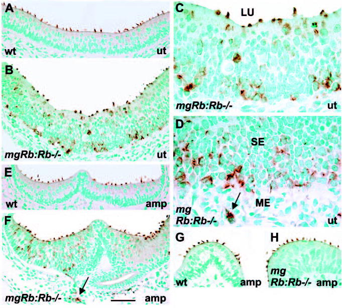
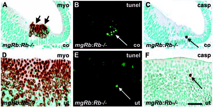
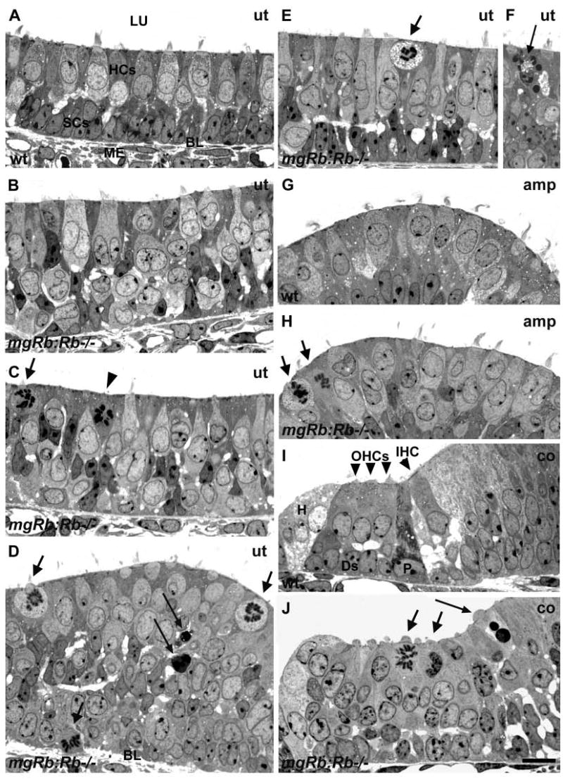
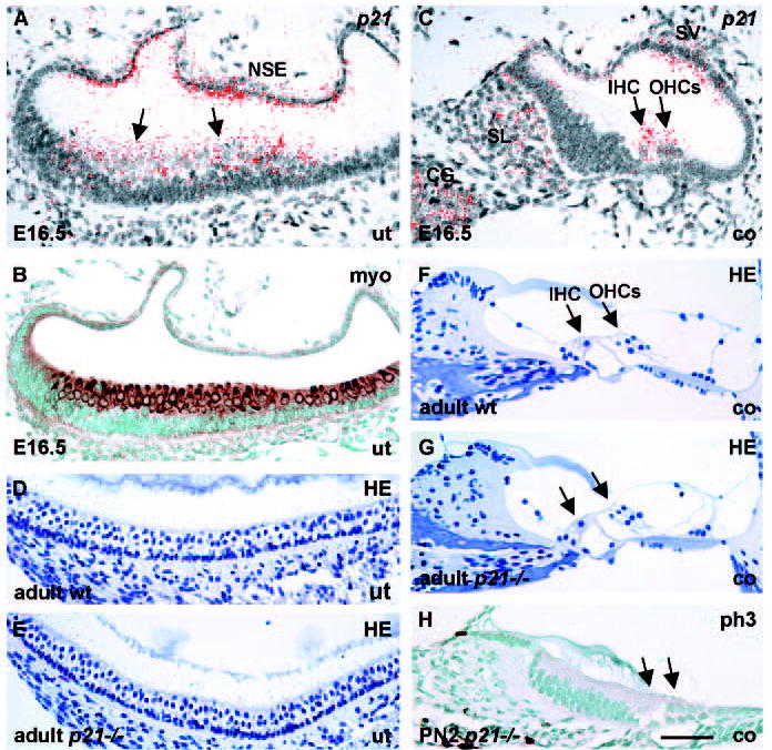
Precursors of cochlear and vestibular hair cells of the inner ear exit the cell cycle at midgestation. Hair cells are mitotically quiescent during late-embryonic differentiation stages and postnatally. We show here that the retinoblastoma gene Rb and the encoded protein pRb are expressed in differentiating and mature hair cells. In addition to Rb, the cyclin dependent kinase inhibitor (CKI) p21 is expressed in developing hair cells, suggesting that p21 is an upstream effector of pRb activity. p21 apparently cooperates with other CKIs, as p21-null mice exhibited an unaltered inner ear phenotype. By contrast, Rb inactivation led to aberrant hair cell proliferation, as analysed at birth in a loss-of-function/transgenic mouse model. Supernumerary hair cells expressed various cell type-specific differentiation markers, including components of stereocilia. The extent of alterations in stereociliary bundle morphology ranged from near-normal to severe disorganization. Apoptosis contributed to the mutant phenotype, but did not compensate for the production of supernumerary hair cells, resulting in hyperplastic sensory epithelia. The Rb-null-mediated proliferation led to a distinct pathological phenotype, including multinucleated and enlarged hair cells, and infiltration of hair cells into the mesenchyme. Our findings demonstrate that the pRb pathway is required for hair cell quiescence and that manipulation of the cell cycle machinery disrupts the coordinated development within the inner ear sensory epithelia.


Figures

Fig. 1
Fig. 1
pRb expression in hair cells of the inner ear. pRb immunostaining; Rb in situ hybridization. (A,B) pRb staining in the early-and later-differentiating HCs of the embryonic saccule. (C,D) pRb and Rb expressions in HCs of the late-embryonic utricle. (E) A subpopulation of HCs is pRb-positive in the adult utricle. (F,G) pRb expression in inner and outer HCs of the cochlea at birth and early postnatal life. In addition to HCs, the greater epithelial ridge shows weak expression at birth. (H) In the mature organ of Corti, pRb expression is detected only in inner HCs. Supporting cells are negative at all stages. (I,J) pRb is not expressed in the hyperplastic sensory epithelium of utricle and cochlea of mgRb:Rb−/− mutants at E18.5. Abbreviations: wt, wild type; sac, saccule; ut, utricle; co, cochlea; IHC, inner hair cell; OHCs, outer hair cells; GER, greater epithelial ridge; HCs, hair cells; SCs, supporting cells. Arrows in G,H,J indicate HCs. Scale bar: 70 μm.
Fig. 2
Fig. 2
Proliferation and differentiation in the cochleas of mgRb:Rb–/– embryos. Myosin VI-and phospho-histone H3 immunostaining; Math1 and Fgf10 in situ hybridization. (A,A′) At birth, myosin VI is expressed in inner and outer HCs of control cochleas and also in supernumerary HCs of mutants. (B-C′) At late-embryogenesis, p27- and p75NTR-positive supporting cell populations (arrows) are seen both in controls and mutants. (D) A myosin VI-stained mitotic HC (arrow) in a mutant cochlea at birth. (E,E′) At birth, phospho-histone H3 staining shows ectopic mitoses in the greater epithelial ridge and the region of inner and outer HCs in mutant cochleas. These are not present in controls. (E″) Most of these mitotic HCs with strongly stained condensed chromatin are located near the lumen. (F,F′) At E14.5, phospho-histone H3- stained cells are seen in the caudal wall of the cochlea of both mutants and controls. (G,G′) At birth, Math1 is expressed in cochlear HCs of controls and also in supernumerary HCs of mutants. (H,H′) At birth, Fgf10 is expressed in the greater epithelial ridge of control cochleas and in the thickened ridge of mutants. Abbreviations: wt, wild type; ph3, phospho-histone H3; myo, myosin VI; GER, greater epithelial ridge; IHC, inner hair cell; OHCs, outer hair cells; CN, cochlear nerve. Scale bar: 70 μm for A-C′,E-F′); 30 μm for D; 80 μm for G-H′).
Fig. 3
Fig. 3
Mitosis and differentiation of vestibular hair cells of mgRb:Rb−/− embryos. Myosin VI- and phospho-histone H3- immunostaining. (A,A′) When compared with controls at birth, utricular sensory epithelia of mutants are hyperplastic because of an excess of myosinVI-positive HCs, some of which have penetrated into the mesenchyme (arrows). (B,B′) In contrast to the saccular sensory epithelium of controls in which HCs occupy the lumenal layer, HCs are distributed throughout the epithelium in mutants. (C,C′) In contrast to controls, ampullary sensory epithelia of mutants are hyperplastic because of overproduction of myosin VI-positive HCs, some of which invade the mesenchyme (arrow). (D) An oblique section through the utricular sensory epithelium of a mutant shows mitotic figures (arrows) in myosin VI-stained HCs. (E) A cross-section through the utricle shows that mitotic HCs (arrows) have rounded shape and occupy both lumenal and deeper epithelial layers. (F-H′) Phospho-histone H3- stained utricular (F,F′), saccular (G,G′) and ampullary (H,H′) sensory epithelia at birth show higher numbers of mitotic cells in mutants when compared with controls. (I,I′) At E14.5, early-differentiating saccular HCs of mutants undergo mitoses, in contrast to controls. Differentiating HCs occupy the lumenal layer and have a large nucleus. Abbreviations: wt, wild type; myo, myosin VI; ph3, phospho-histone H3. Scale bar: 120 μm for A,A′,F,F′); 70 μm for B-C′,G-I′; 30 μm for D,E.
Fig. 4
Fig. 4
Stereociliary bundles of vestibular hair cells of mgRb:Rb−/− mutants at birth, as shown by espin-immunostaining. (A) Espin is localized to HC stereocilia of control utricules. (B) In mutants, espin-positive HCs are situated throughout the thickened utricular sensory epithelium. Several HCs at the surface show abnormal bundles. (C,D) High magnification views show HCs with aberrant stereocilia in the utricle and in the adjacent mesenchyme (arrow) of mutants. (E,F) In contrast to controls, mutants show espin-stained HCs at different levels of the ampullary sensory epithelium. HCs in the mesenchyme are also espin positive (arrow). (G,H) In addition to the distinct bundle abnormalities, stereociliary bundle morphologies comparable with controls are seen in some HCs of mutants, especially in ampullae. Abbreviations: wt, wild type; ut; utricle; amp, ampulla; SE, sensory epithelium; ME, mesenchyme; LU, lumen. Scale bar: 70 μm for A-F; 60 μm for G,H; 30 μm for C,D.
Fig. 5
Fig. 5
Apoptosis in the inner ear of mgRb:Rb−/− mutants at E18.5. (A-C) In a mutant cochlea, the myosin VI-stained region of supernumerary HCs, which comprises mitotic figures (A, short arrows), shows TUNEL- (B) and cleaved caspase 3- (C) positive cells. Views are from adjacent sections. Compare with normal morphology in Fig. 2A. (D-F) TUNEL- and caspase 3-positive cells are present in the myosin VI-stained region of a hyperplastic utricular sensory epithelium. Compare with normal morphology in Fig. 3A. Long arrows indicate apoptotic profiles. Abbreviations: co, cochlea; ut, utricle; myo, myosin VI; casp, cleaved caspase 3. Scale bar: 60 μm.
Fig. 6
Fig. 6
Mitosis, apoptosis and multinucleation of hair cells of mgRb:Rb/ mutants at E18.5, as shown in semi-thin sections. (A) Normal utricular sensory epithelium shows HCs at the lumenal surface and underlying supporting cells. (B) A mutant utricle shows disorganized cell layering and HCs with an atypical nucleus or with two or more nuclei. Some of these HCs show near-normal stereociliary bundles. Some HCs seem to extend from the surface deep into the epithelium. (C) Mitotic cells with rounded morphology and nascent stereocilia (arrow) at the lumenal surface are intermixed with HCs with aberrant nuclear morphologies. A putative supporting cell that has translocated to the surface for mitosis is marked by an arrowhead. (D) Mitotic HCs (short arrows) are found mostly at the lumenal surface, but also in deeper layers, such as in this view where a cell undergoing mitosis is penetrating through the basal lamina. Scattered apoptotic profiles are also found (long arrows). (E,F) A mitotic cell (short arrow) showing signs of degeneration in the cytoplasm. Mitotic cells occasionally showed apoptotic figures (long arrow). (G,H) In contrast to controls, the ampullary sensory epithelia of mutants contain mitotic cells (arrows). In addition, several HCs with abnormal nuclei are found and many of them have stereociliary bundles with a morphology comparable with controls. (I,J) A transverse view through the organ of Corti of a normal cochlea shows four HCs and the underlying supporting cells. In the region of organ of Corti of mutants, high numbers of nuclei are seen, some of them having mitotic figures (short arrows) and a few apoptotic profiles (long arrow). Abbreviations: wt, wild type; ut, utricle; amp, ampulla; co, cochlea; LU, lumen, BL, basal lamina; ME, mesenchyme; HCs, hair cells; SCs, supporting cells; IHC, inner hair cell; OHCs, outer hair cells; P, pillar cell; Ds, Deiters’ cell, H, Hensen’s cell. Scale bar: 15 μm for A-J.
Fig. 7
Fig. 7
p21 expression in the developing inner ear and the phenotype of p21 knockout mice. p21 in situ hybridization; myosin VI and phospho-histone H3 immunostaining; Haematoxylin staining. (A,B) As shown in adjacent sections, p21 and myosin VI are expressed in utricular HCs (arrows) at E16.5. p21 is also found in the utricular non-sensory epithelium. (C) In the cochlea at E16.5, p21 is expressed in HCs and in stria vascularis, spiral limbus and cochlear ganglion neurons. (D,E) Histology of the utricular sensory epithelium of adult p21−/− mice is indistinguishable from controls, as shown by Haematoxylin staining. (F,G) Histology of the organ of Corti of adult p21 knockouts is comparable with controls. (H) Mitoses are not induced in the cochleas of p21-null mutants, as shown at P2 by phospho-histone H3-staining (compare with control in Fig. 2E). Arrows in G,H indicate HCs. Abbreviations: wt, wild type; ut, utricle; co, cochlea; HE, Haematoxylin; SL, spiral limbus; CG, cochlear ganglion; SV, stria vascularis; IHC, inner hair cell; OHCs, outer hair cells; NSE, non-sensory epithelium; myo, myosin VI; ph3, phospho-histone H3. Scale bar: 70 μm for A-C,H; 80 μm for D-G.

Similar articles

Cited by

References

    1. Bermingham NA, Hassan BA, Price SD, Vollrath MA, Ben-Arie N, Eatock RA, Bellen HJ, Lysakowski A, Zoghbi HY. Math1: an essential gene for the generation of inner ear hair cells. Science. 1999;284:1837–1841. - PubMed
    1. Bhave SA, Stone JS, Rubel EW, Coltrera MD. Cell cycle progression in gentamicin-damaged avian cochleas. J Neurosci. 1995;16:4618–4628. - PMC - PubMed
    1. Brugarolas J, Chandrasekaran C, Gordon JI, Beach D, Jacks T, Hannon GJ. Radiation-induced cell cycle arrest compromised by p21 deficiency. Nature. 1995;377:552–557. - PubMed
    1. Chau BN, Wang JY. Coordinated regulation of life and death by RB. Nat Rev Cancer. 2003;3:130–138. - PubMed
    1. Chen PL, Riley DJ, Chen Y, Lee WH. Retinoblastoma protein positively regulates terminal adipocyte differentiation through direct interaction with C/EBPs. Genes Dev. 1996;10:2794–2804. - PubMed

Publication types

Substances