. 2005 Jul;16(7):3411-24.
doi: 10.1091/mbc.e04-11-1009. Epub 2005 Apr 20.

Lamin A/C-dependent localization of Nesprin-2, a giant scaffolder at the nuclear envelope

Affiliations

Lamin A/C-dependent localization of Nesprin-2, a giant scaffolder at the nuclear envelope

Thorsten Libotte et al. Mol Biol Cell. 2005 Jul.

Abstract

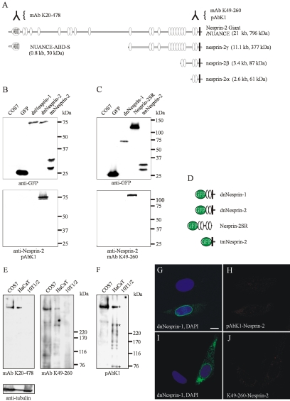
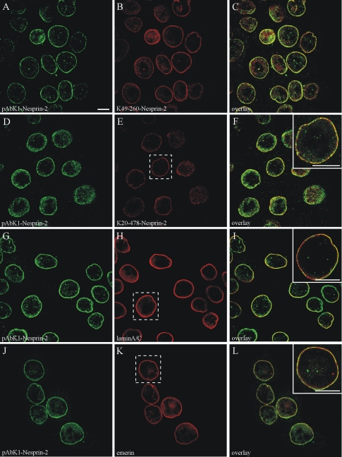
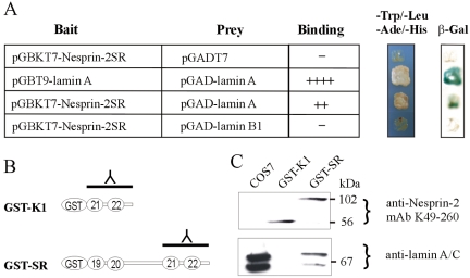
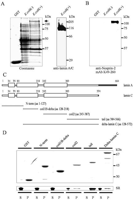
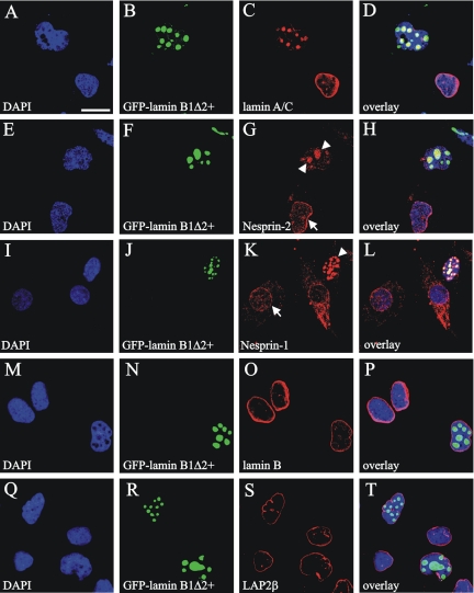
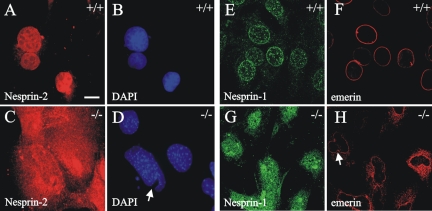
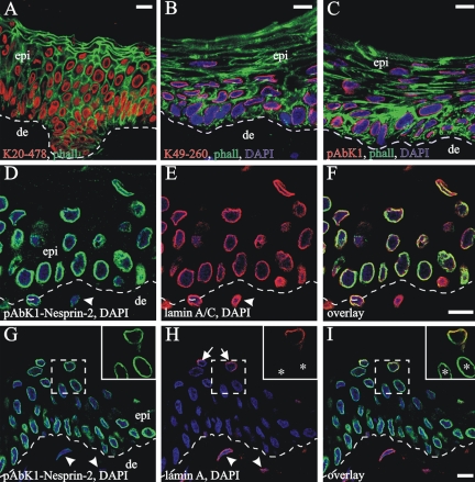
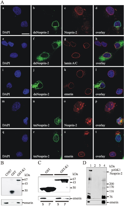
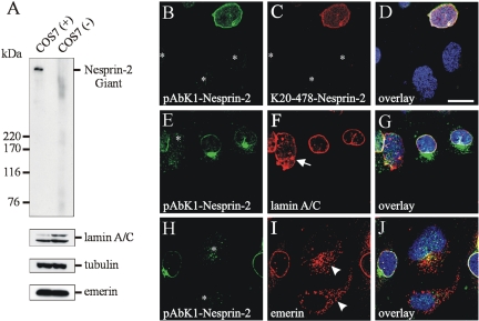
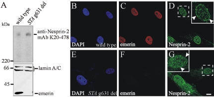
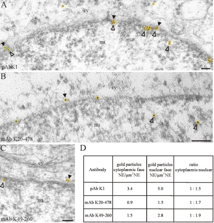
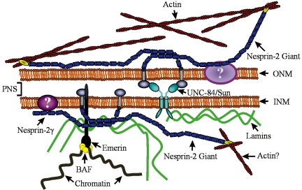
The vertebrate proteins Nesprin-1 and Nesprin-2 (also referred to as Enaptin and NUANCE) together with ANC-1 of Caenorhabditis elegans and MSP-300 of Drosophila melanogaster belong to a novel family of alpha-actinin type actin-binding proteins residing at the nuclear membrane. Using biochemical techniques, we demonstrate that Nesprin-2 binds directly to emerin and the C-terminal common region of lamin A/C. Selective disruption of the lamin A/C network in COS7 cells, using a dominant negative lamin B mutant, resulted in the redistribution of Nesprin-2. Furthermore, using lamin A/C knockout fibroblasts we show that lamin A/C is necessary for the nuclear envelope localization of Nesprin-2. In normal skin where lamin A/C is differentially expressed, strong Nesprin-2 expression was found in all epidermal layers, including the basal layer where only lamin C is present. This indicates that lamin C is sufficient for proper Nesprin-2 localization at the nuclear envelope. Expression of dominant negative Nesprin-2 constructs and knockdown studies in COS7 cells revealed that the presence of Nesprin-2 at the nuclear envelope is necessary for the proper localization of emerin. Our data imply a scaffolding function of Nesprin-2 at the nuclear membrane and suggest a potential involvement of this multi-isomeric protein in human disease.


Figures

Figure 1.
Figure 1.
Splice variants of the Nesprin-2 locus and characterization of novel C-terminal Nesprin-2 antibodies. (A) Structural features of known Nesprin-2 isoforms. Nesprin-2 Giant/NUANCE consists of an N-terminal actin-binding domain (ABD), followed by several spectrin repeats (ovals) and a C-terminal transmembrane domain (black bar). Epitopes and the identity of various Nesprin-2 antibodies used in this report are indicated (inverted Y). (B and C) COS7 cells were transiently transfected with various C-terminal Nesprin GFP-fusion proteins. The COS7 cell lysates were subjected to Western blot analysis using anti-GFP–specific monoclonal antibodies to demonstrate expression of fusion proteins (top panels in B and C). Bottom panels in B and C, immunoblot analysis of the same COS7 lysates using the newly generated C-terminal polyclonal (pAb K1) and the mouse monoclonal Nesprin-2 (mAb K49-260) antibodies, verifies their specificity to the Nesprin-2 protein. (D) Schematic diagram of the GFP constructs used in this study. (E and F) COS7, HaCaT, and mouse embryonic C3H/10T1/2 cell homogenates were separated on 3–15% gradient SDS-polyacrylamide gels and blotted (Padmakumar et al., 2004). Western blot analysis using the N-terminus specific Nesprin-2 antibody mAb K20-478 and the newly generated C-terminal Nesprin-2 antibodies mAb K49–260 and pAbK1, demonstrate the presence of the large 800-kDa Nesprin-2 isoform in COS7 and HaCaT cells. Nesprin-2 expression was not detected in C3H/10T1/2 fibroblast lysates. The blots shown in E originate from the same gel. The same blot was reprobed with anti-β-tubulin antibodies to illustrate equal loading. (G–J) C3H/10T1/2 fibroblasts cells transfected with the dnNesprin-1 GFP fusion protein were subjected to indirect immunofluorescence using the C-terminal Nesprin-2 antibodies pAbK1 and mAb K49–260. DAPI was used to stain nuclei. Images were taken by confocal microscopy. Bar, 10 μm.
Figure 2.
Figure 2.
Nesprin-2 colocalizes with lamin A/C and emerin at the nuclear envelope of HaCaT cells. Immunolocalization studies of HaCaT cells by using anti-Nesprin-2, antilamin A/C, and anti-emerin antibodies. (A–F) HaCaT cells subjected to immunofluorescence using three different Nesprin-2 antibodies (indicated in the bottom left of each frame) demonstrate a similar nuclear localization pattern. Nesprin-2 colocalizes with lamin A/C (G–I) and emerin (J–L) at the nuclear envelope. Note the appearance of yellow in the overlay panels. Insets are higher magnifications of areas in dotted white boxes. DAPI was used to stain nuclei. Images were taken by confocal microscopy. All scale bars, 10 μm.
Figure 3.
Figure 3.
(A) The Nesprin-2SR fragment (aa 6146–6799) was fused to the Gal4 DNA-binding domain, whereas lamin A and B1 sequences were linked to the Gal4-activating domain. The corresponding plasmids were cotransformed into yeast cells and the interactions were assessed by the filter lift β-galactosidase assay. ++++, high blue color development; ++, weak blue color development; –, no blue color development. The growth of the yeast cells on selection media is indicated as well as β-galactosidase stainings of the colony filter lifts. GST pulldown of lamin A/C and emerin by a C-terminal Nesprin-2 fragment. (B) Schematic overview of the fusion proteins used for the GST pulldown assay of COS7 homogenates. GST-K1 consists of the last two spectrin repeats, whereas GST-SR consists of the last four spectrin repeats of Nesprin-2 Giant. The position of the epitope of the C-terminal Nesprin-2 antibody is indicated (inverted Y). (C) Immunoblotting using the C-terminus–specific Nesprin-2 mAb K49-260 demonstrates that equal amounts of the GST fusion proteins were used (top panel). COS7 cell lysates were incubated with the immobilized GST-fusion proteins. The bound proteins were subjected to SDS-PAGE followed by Western blot analysis using lamin A/C–specific antibodies (bottom panel). As a control we used a COS7 total cell lysate.
Figure 4.
Figure 4.
Nesprin-2 interacts with lamin A/C in vitro. (A) Coomassie blue–stained gel containing GST and equal amounts of uninduced (–) and induced (+) E. coli cell lysates expressing GST-lamin A (full-length, arrow). The expression of GST-lamin A was verified by Western blot analysis using lamin A/C (JoL2) antibodies (left panel). The proteins were blotted onto a membrane and subsequently used for blot overlay assays. (B) Blot overlay assay using the GST-SR fusion protein as a probe, followed by immunodetection with mAb K49–260 Nesprin-2 antibody. (C) Schematic overview of the domain architecture of lamin A/C and the GST-lamin C fusion proteins used for the in vitro GST pulldown assays. The unique C-terminal sequences in the lamin A and lamin C isoforms are shaded. (D) Approximately equal amounts of GST and GST lamin C fusion proteins were immobilized on agarose beads and incubated with the Nesprin-2SR recombinant protein. Samples were centrifuged and supernatant (S), pellet (P) fractions were subjected to SDS-PAGE, followed by Coomassie blue staining (top panel) to detect the GST fusion proteins and by silver staining to detect the Nesprin-2SR recombinant protein (bottom panel).
Figure 5.
Figure 5.
Nesprin-2 distribution is lamin A/C dependent. (A–T) COS7 cells were transiently transfected with a plasmid coding for the dominant negative GFP-lamin B1Δ2+ protein and subjected to immunofluorescence. Lamin A/C is redistributed into the GFP-positive nuclear patches in transfected cells and is no longer prominent at the nuclear membrane (C). Nesprin-2 (recognized by mAb K20-478) redistributes also into these GFP positive patches (G; arrowheads), whereas the endogenous protein is localized at the nuclear membrane in nontransfected cells (G, arrow). A similar pattern is also observed for Nesprin-1 (I–L). In untransfected cells Nesprin-1 is being found at the nuclear membrane (K; arrows), whereas in transfected cells Nesprin-1 colocalizes with the intranuclear GFP-patches (K; arrowheads). Endogenous lamin B and LAP2β were found to be unaffected in transfected cells and were retained at the nuclear rim (O and S, respectively). DNA was stained with DAPI. Analysis was done by confocal microscopy. Images were merged to visualize colocalization (D, H, L, P, and T). Bar, 25 μm.
Figure 6.
Figure 6.
The distribution of Nesprins is affected in cells that do not express lamins A/C. (A–H) Wild-type and knockout lamin A/C fibroblasts were subjected to immunofluorescence using Nesprin-2 (pAbK1; A and C), Nesprin-1 (E and G, using a C-terminal Nesprin polyclonal antibody) and emerin (F and H) antibodies. Both Nesprin-2 (C) as well as Nesprin-1 (G) no longer localize at the nuclear membrane in lamin A/C knockout cells. Notice the confined nuclear envelope staining of Nesprin-2 (A) and Nesprin-1 (E) in wild-type cells. Nuclei deformations of the lamin A/C mutants are denoted by arrows. DNA was stained with DAPI. Images were taken by confocal microscopy except for A–D, which were obtained by conventional microscopy. Bar, 10 μm.
Figure 7.
Figure 7.
In human skin, lamin C is sufficient for the proper nuclear localization of Nesprin-2. (A–C) Human frozen skin sections were stained with N- and C-terminally directed Nesprin-2 antibodies as indicated in the lower left of each frame and FITC-conjugated phalloidin. Note that the antibodies share an identical nuclear staining pattern and detect Nesprin-2 throughout the living layers of the epidermis. (D–I) Human skin sections counterstained with anti-Nesprin-2- (pAbK1; D and G), JoL2- (anti-lamin A/C; E), and JoL4- (anti-lamin A; H) specific antibodies. Insets are higher magnifications of areas in dotted white boxes. Lamin A–negative keratinocytes are indicated by asterisks in the insets. Arrows indicate lamin A–positive suprabasal keratinocytes, whereas arrowheads indicate lamin A–positive dermal fibroblasts (H). Dermal fibroblasts are indicated by arrowheads; notice the reduced Nesprin-2 staining in the dermal fibroblasts (D and G; arrowheads). The dotted line denotes the basement membrane, which was visualized by counterstaining with a6β4 antibodies and a Cy5-conjugated secondary antibody. DNA was stained with DAPI Images were taken by confocal microscopy. epi, epidermis; de, dermis. All scale bars, 10 μm.
Figure 8.
Figure 8.
Dominant negative interference with the Nesprin-2 localization in COS7 cells influences the localization of emerin. (A) COS7 cells were transiently transfected with plasmids allowing expression of the GFP-tagged C-terminal Nesprin-2 dominant negative polypeptides dnNesprin-2 (a–l) and tmNesprin-2 (m–t). The constructs used are described in Material and Methods and a schematic overview of their composition is shown in Figure 1D. Immunofluorescence studies were performed using Nesprin-2 (c and o), lamin A/C (g), and emerin antibodies (k and s). Although both constructs localize to the nuclear envelope and displace the endogenous Nesprin-2 proteins (c and o), a mislocalization of emerin in the cytoplasm is observed only for the tmNesprin-2 GFP construct (s). The cells were fixed with paraformaldehyde. DNA was stained with DAPI. Analysis was done by confocal microscopy. Images were merged to visualize colocalization (d, h, l, p, and t). Bar, 25 μm. (B) Emerin associates directly with Nesprin-2 in vitro. GST and GST-K1 fusion proteins were immobilized on glutathione agarose beads and were incubated either with COS7 cell lysates (B) or with recombinant emerin protein (C). Samples were centrifuged and supernatant (S) and pellet (P) fractions were subjected to SDS-PAGE, followed by immunoblot analysis to detect the GST fusions proteins (top panels) and emerin (bottom panels). (D) CoIP of Nesprin-2 and emerin from HaCaT cell lysates. HaCaT cells were lysed, and the supernatants were incubated with anti-Nesprin-2 (mAb K49-260) antibodies. The immunocomplexes were analyzed by SDS-PAGE and subjected to immunoblotting with anti-Nesprin-2 (pAbK1) and emerin antibody. Lane 1, total cell lysate; lane 2, lysate precipitated with Protein A Sepharose beads; lane 3, lysate precipitated with anti GFP antibody on Protein A Sepharose beads; lane 4, lysate precipitated with anti-Nesprin-2 antibody on Protein A/G Sepharose beads.
Figure 9.
Figure 9.
The emerin distribution is affected in cells where Nesprin-2 expression has been silenced by siRNA. (A) COS7 cells were transiently transfected with the pSUPER plasmid expressing RNA duplexes against the N-terminus of Nesprin-2. Cell lysates were subjected to Western blot analysis using Nesprin-2 (pAbK1), lamin A/C, emerin and tubulin antibodies, illustrating the efficiency of the Nesprin-2 knockdown. The distribution of Nesprin-2 (B, E, and H; using pAbK1 and C; using K20-478), lamin A/C (F), and emerin (I) was investigated by indirect immunofluorescence in knockdown Nesprin-2 COS7 cells (indicated by an asterisk). In knockdown Nesprin-2 cells, lamin A/C remained at the nuclear envelope (F, arrow), whereas emerin was dispersed in cytoplasmic aggregates (I; arrowheads). Cells were fixed with paraformaldehyde. DNA was stained with DAPI. Analysis was done by confocal microscopy. Images were merged to visualize colocalization (D, G, and J). Bar, 25 μm.
Figure 10.
Figure 10.
Emerin is not necessary for the NE localization of Nesprin-2 in dermal fibroblasts. (A) Immunoblot analysis of wild-type and emerin-deficient (STA g631 del) human fibroblasts lysates using anti-Nesprin-2 (mAb K20-478), lamin A/C- and emerin-specific antibodies. Wild-type (B–D) and emerin-deficient cells (E–G) were subjected to immunofluorescence using antibodies against Nesprin-2 (mAb K20-478) and emerin. Insets are higher magnifications of areas in dotted white boxes. Arrowheads denote the Nesprin-2 staining at the nuclear envelope. Note the persistence of the nuclear staining pattern of Nesprin-2 in the mutant cells (G). The images were taken by confocal microscopy. Nuclei were visualized by DAPI staining. Bar, 10 μm.
Figure 11.
Figure 11.
Immunogold EM examination of Nesprin-2. In HaCaT cells pAbK1 (A), mAb K20-478 N-terminal (B) and mAb K49–20 C-terminal Nesprin-2–specific antibodies (C) recognize nuclear (open arrowheads) as well as cytoplasmic (black arrows) Nesprin-2 protein at the NE. The gold particles appear often in clusters (A) and are marked with yellow circles. Quantification of Nesprin-2 antibody labeling at the nuclear envelope (D). nu, nucleus; cy, cytoplasm. All scale bars, 100 nm.
Figure 12.
Figure 12.
A model, illustrating the interactions of Nesprin-2 Giant and its isoforms (Nesprin-2γ) with various cytoplasmic and nucleoplasmic components at the nuclear membrane (see Discussion for details). Unknown complexes, which may include peripheral (members of the 4.1 protein family) as well as integral elements of the nuclear membrane are depicted as large ovals. ONM, outer nuclear membrane; INM, inner nuclear membrane; PNS, perinuclear space; BAF, barrier-to-autointegration factor.

Similar articles

Cited by

References

    1. Apel, E. D., Lewis, R. M., Grady, R. M., and Sanes, J. R. (2000). Syne-1, a dystrophin- and Klarsicht-related protein associated with synaptic nuclei at the neuromuscular junction. J. Biol. Chem. 275, 31986–31995. - PubMed
    1. Bengtsson, L., and Wilson, K. L. (2004). Multiple and surprising new functions for emerin, a nuclear membrane protein. Curr. Opin. Cell Biol. 16, 1–7. - PubMed
    1. Burke, B., and Stewart, C. L. (2002). Life at the edge: the nuclear envelope and human disease. Nat. Rev. Mol. Cell. Biol. 3, 575–585. - PubMed
    1. Dreuillet, C., Tillit, J., Kress, M., and Ernoult-Lange, M. (2002). In vivo and in vitro interaction between human transcription factor MOK2 and nuclear lamin A/C. Nucleic Acids Res. 30, 4634–4642. - PMC - PubMed
    1. Ellis, D. J., Jenkins, H. E., Whitfield, W.G.F., and Hutchison, C. J. (1997). GST-lamin fusion proteins act as dominant negative mutants in Xenopus egg extract and reveal the function of the lamina in DNA replication. J. Cell Sci. 110, 2507–2518. - PubMed

Publication types

MeSH terms