Mechanical strain opens connexin 43 hemichannels in osteocytes: a novel mechanism for the release of prostaglandin
- PMID: 15843434
- PMCID: PMC1165395
- DOI: 10.1091/mbc.e04-10-0912
Mechanical strain opens connexin 43 hemichannels in osteocytes: a novel mechanism for the release of prostaglandin
Abstract
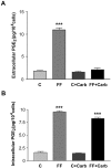
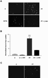
Mechanosensing bone osteocytes express large amounts of connexin (Cx)43, the component of gap junctions; yet, gap junctions are only active at the small tips of their dendritic processes, suggesting another function for Cx43. Both primary osteocytes and the osteocyte-like MLO-Y4 cells respond to fluid flow shear stress by releasing intracellular prostaglandin E2 (PGE2). Cells plated at lower densities release more PGE2 than cells plated at higher densities. This response was significantly reduced by antisense to Cx43 and by the gap junction and hemichannel inhibitors 18 beta-glycyrrhetinic acid and carbenoxolone, even in cells without physical contact, suggesting the involvement of Cx43-hemichannels. Inhibitors of other channels, such as the purinergic receptor P2X7 and the prostaglandin transporter PGT, had no effect on PGE2 release. Cell surface biotinylation analysis showed that surface expression of Cx43 was increased by shear stress. Together, these results suggest fluid flow shear stress induces the translocation of Cx43 to the membrane surface and that unapposed hemichannels formed by Cx43 serve as a novel portal for the release of PGE2 in response to mechanical strain.
Figures


Similar articles
-
Hemichannels formed by connexin 43 play an important role in the release of prostaglandin E(2) by osteocytes in response to mechanical strain.Cell Commun Adhes. 2003 Jul-Dec;10(4-6):259-64. doi: 10.1080/cac.10.4-6.259.264. Cell Commun Adhes. 2003. PMID: 14681026
-
Effects of mechanical strain on the function of Gap junctions in osteocytes are mediated through the prostaglandin EP2 receptor.J Biol Chem. 2003 Oct 31;278(44):43146-56. doi: 10.1074/jbc.M302993200. Epub 2003 Aug 25. J Biol Chem. 2003. PMID: 12939279
-
Expression of functional gap junctions and regulation by fluid flow in osteocyte-like MLO-Y4 cells.J Bone Miner Res. 2001 Feb;16(2):249-59. doi: 10.1359/jbmr.2001.16.2.249. J Bone Miner Res. 2001. PMID: 11204425
-
The Role of Connexin Channels in the Response of Mechanical Loading and Unloading of Bone.Int J Mol Sci. 2020 Feb 9;21(3):1146. doi: 10.3390/ijms21031146. Int J Mol Sci. 2020. PMID: 32050469 Free PMC article. Review.
-
Connexin 43 hemichannels and prostaglandin E2 release in anabolic function of the skeletal tissue to mechanical stimulation.Front Cell Dev Biol. 2023 Apr 13;11:1151838. doi: 10.3389/fcell.2023.1151838. eCollection 2023. Front Cell Dev Biol. 2023. PMID: 37123401 Free PMC article. Review.
Cited by
-
Primary Osteocyte Supernatants Metabolomic Profiling of Two Transgenic Mice With Connexin43 Dominant Negative Mutants.Front Endocrinol (Lausanne). 2021 May 18;12:649994. doi: 10.3389/fendo.2021.649994. eCollection 2021. Front Endocrinol (Lausanne). 2021. PMID: 34093433 Free PMC article.
-
Blockage of hemichannels alters gene expression in osteocytes in a high magneto-gravitational environment.Front Biosci (Landmark Ed). 2017 Jan 1;22(5):783-794. doi: 10.2741/4516. Front Biosci (Landmark Ed). 2017. PMID: 27814646 Free PMC article.
-
The gap junction cellular internet: connexin hemichannels enter the signalling limelight.Biochem J. 2006 Jul 1;397(1):1-14. doi: 10.1042/BJ20060175. Biochem J. 2006. PMID: 16761954 Free PMC article. Review.
-
Spinning around or stagnation - what do osteoblasts and chondroblasts really like?Eur J Med Res. 2010 Jan 29;15(1):35-43. doi: 10.1186/2047-783x-15-1-35. Eur J Med Res. 2010. PMID: 20159670 Free PMC article.
-
Functional Roles of Connexins and Gap Junctions in Osteo-Chondral Cellular Components.Int J Mol Sci. 2023 Feb 19;24(4):4156. doi: 10.3390/ijms24044156. Int J Mol Sci. 2023. PMID: 36835567 Free PMC article. Review.
References
-
- Aarden, E. M., Burger, E. H., and Nijweide, P. J. (1994). Function of osteocytes in bone. J. Cell. Biochem. 55, 287–299. - PubMed
-
- Ajubi, N. E., Klein-Nulend, J., Nijweide, P. J., Vrijheid-lammers, T., Alblas, M. J., and Burger, E. H. (1996). Pulsating fluid flow increases prostaglandin production by cultured chicken osteocytes-a cytoskeleton-dependent process. Biochem. Biophys. Res. Commun. 225, 62–68. - PubMed
-
- Baylink, T. M., Mohan, S., Fitzsimmons, R. J., and Baylink, D. J. (1995). Evidence that the mitogenic effect of prostaglandin E2 on human bone cells involves protein kinase C and calcium pathways. J. Bone Miner. Res. 9 (suppl 31), B23.
-
- Baylink, T. M., Mohan, S., Fitzsimmons, R. J., and Baylink, D. J. (1996). Evaluation of signal transduction mechanisms for the mitogenic effects of prostaglandin E2 in normal human bone cells in vitro. J. Bone Miner. Res. 11, 1413–1418. - PubMed
Publication types
MeSH terms
Substances
Grants and funding
LinkOut - more resources
Full Text Sources
Other Literature Sources
Molecular Biology Databases
Miscellaneous

