

Role of matrix extracellular phosphoglycoprotein in the pathogenesis of X-linked hypophosphatemia
- PMID: 15843468
- PMCID: PMC1484502
- DOI: 10.1681/ASN.2004121060
Role of matrix extracellular phosphoglycoprotein in the pathogenesis of X-linked hypophosphatemia
Abstract
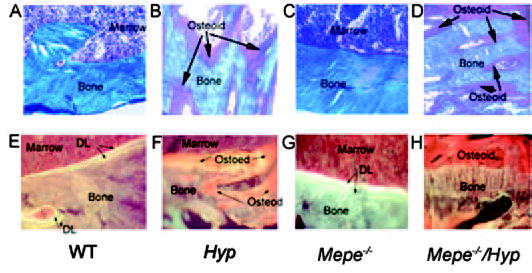
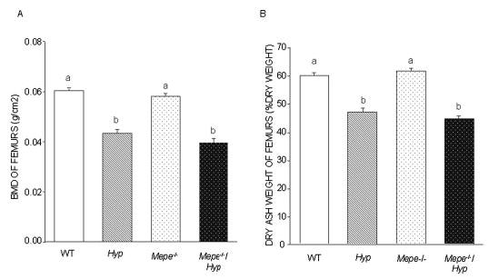
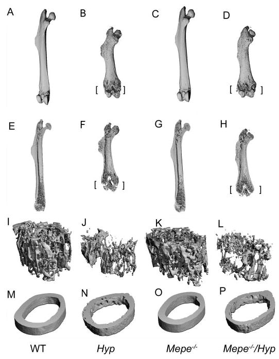
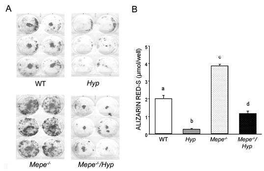
X-linked hypophosphatemia (XLH), a disorder characterized by hypophosphatemia, impaired skeletal mineralization, and aberrant regulation of 1, 25(OH)(2)D(3), is caused by inactivating mutations of Phex, which results in the accumulation of putative phosphaturic factors, called phosphatonins. Matrix extracellular phosphoglycoprotein (Mepe) is a proposed candidate for phosphatonin. The authors found that Hyp mice had increased expression of the MEPE and another phosphaturic factor, Fgf23. To establish MEPE's role in the pathogenesis of the XLH, Mepe-deficient mice were back-crossed onto the Hyp mouse homologue of XLH and phenotypes of wild-type, Mepe(-/-), Hyp, and Mepe(-/-)/Hyp mice were examined. Transfer of Mepe deficiency onto the Phex-deficient Hyp mouse background failed to correct hypophosphatemia and aberrant serum 1,25(OH)(2)D(3) levels. Increased Fgf23 levels in Hyp mice were not affected by superimposed Mepe deficiency. In addition, Mepe-deficient Hyp mice retained bone mineralization defects in vivo, characterized by decreased bone mineral density, reduced mineralized trabecular bone volume, lower flexural strength, and histologic evidence of osteomalacia; however, cultures of Hyp-derived bone marrow stromal cells in the absence of Mepe showed improved mineralization and normalization of osteoblast gene expression profiles observed in cells derived from Mepe-null mice. These results demonstrate that MEPE elevation in Hyp mice does not contribute to the hypophosphatemia associated with inactivating Phex mutations and is therefore not phosphatonin.
Figures
Similar articles
-
FGF23, PHEX, and MEPE regulation of phosphate homeostasis and skeletal mineralization.Am J Physiol Endocrinol Metab. 2003 Jul;285(1):E1-9. doi: 10.1152/ajpendo.00016.2003. Am J Physiol Endocrinol Metab. 2003. PMID: 12791601 Review.
-
MEPE-ASARM peptides control extracellular matrix mineralization by binding to hydroxyapatite: an inhibition regulated by PHEX cleavage of ASARM.J Bone Miner Res. 2008 Oct;23(10):1638-49. doi: 10.1359/jbmr.080601. J Bone Miner Res. 2008. PMID: 18597632
-
Mepe, the gene encoding a tumor-secreted protein in oncogenic hypophosphatemic osteomalacia, is expressed in bone.Genomics. 2001 Jun 15;74(3):342-51. doi: 10.1006/geno.2001.6553. Genomics. 2001. PMID: 11414762
-
Phosphorylated acidic serine-aspartate-rich MEPE-associated motif peptide from matrix extracellular phosphoglycoprotein inhibits phosphate regulating gene with homologies to endopeptidases on the X-chromosome enzyme activity.J Endocrinol. 2007 Jan;192(1):261-7. doi: 10.1677/joe.1.07059. J Endocrinol. 2007. PMID: 17210763 Free PMC article.
-
The wrickkened pathways of FGF23, MEPE and PHEX.Crit Rev Oral Biol Med. 2004 Sep 1;15(5):264-81. doi: 10.1177/154411130401500503. Crit Rev Oral Biol Med. 2004. PMID: 15470265 Free PMC article. Review.
Cited by
-
Age dependent regulation of bone-mass and renal function by the MEPE ASARM-motif.Bone. 2015 Oct;79:131-42. doi: 10.1016/j.bone.2015.05.030. Epub 2015 Jun 4. Bone. 2015. PMID: 26051469 Free PMC article.
-
Overexpression of the DMP1 C-terminal fragment stimulates FGF23 and exacerbates the hypophosphatemic rickets phenotype in Hyp mice.Mol Endocrinol. 2012 Nov;26(11):1883-95. doi: 10.1210/me.2012-1062. Epub 2012 Aug 28. Mol Endocrinol. 2012. PMID: 22930691 Free PMC article.
-
ASARM mineralization hypothesis: a bridge too far?J Bone Miner Res. 2010 Apr;25(4):692-4. doi: 10.1002/jbmr.69. J Bone Miner Res. 2010. PMID: 20200985 Free PMC article. No abstract available.
-
Matrix extracellular phosphoglycoprotein inhibits phosphate transport.J Am Soc Nephrol. 2008 Dec;19(12):2313-20. doi: 10.1681/ASN.2008030315. Epub 2008 Nov 12. J Am Soc Nephrol. 2008. PMID: 19005008 Free PMC article.
-
Inherited hypophosphatemic disorders in children and the evolving mechanisms of phosphate regulation.Rev Endocr Metab Disord. 2008 Jun;9(2):171-80. doi: 10.1007/s11154-008-9075-3. Epub 2008 Mar 26. Rev Endocr Metab Disord. 2008. PMID: 18365315 Review.
References
-
- The HYP Consortium. A gene (PEX) with homologies to endopeptidases is mutated in patients with X-linked hypophosphatemic rickets. Nat Genet. 1995;11:130–136. - PubMed
-
- Strom TM, Francis F, Lorenz B, Boddrich A, Econs MJ, Lehrach H, Meitinger T. Pex gene deletions in Gy and Hyp mice provide mouse models for X-linked hypophosphatemia. Hum Mol Genet. 1997;6:165–171. - PubMed
-
- Du L, Desbarats M, Viel J, Glorieux FH, Cawthorn C, Ecarot B. cDNA cloning of the murine Pex gene implicated in X-linked hypophosphatemia and evidence for expression in bone. Genomics. 1996;36:22–28. - PubMed
Publication types
MeSH terms
Substances
Grants and funding
LinkOut - more resources
Full Text Sources
Molecular Biology Databases
