. 2005 May 1;174(9):5215-23.
doi: 10.4049/jimmunol.174.9.5215.

TGF-beta 1 attenuates the acquisition and expression of effector function by tumor antigen-specific human memory CD8 T cells

Affiliations

TGF-beta 1 attenuates the acquisition and expression of effector function by tumor antigen-specific human memory CD8 T cells

Mojgan Ahmadzadeh et al. J Immunol. .

Abstract

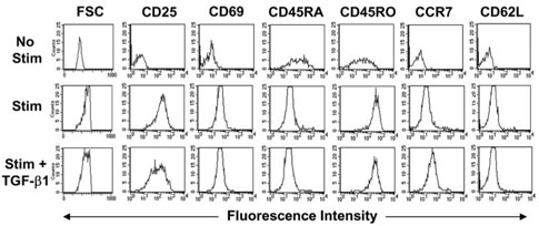
TGF-beta1 is a potent immunoregulatory cytokine. However, its impact on the generation and effector function of Ag-specific human effector memory CD8 T cells had not been evaluated. Using Ag-specific CD8 T cells derived from melanoma patients immunized with the gp100 melanoma Ag, we demonstrate that the addition of TGF-beta1 to the initial Ag activation cultures attenuated the gain of effector function by Ag-specific memory CD8 T cells while the phenotypic changes associated with activation and differentiation into effector memory were comparable to control cultures. These activated memory CD8 T cells consistently expressed lower mRNA levels for T-bet, suggesting a mechanism for TGF-beta1-mediated suppression of gain of effector function in memory T cells. Moreover, TGF-beta1 induced a modest expression of CCR7 on Ag-activated memory CD8 T cells. TGF-beta1 also suppressed cytokine secretion by Ag-specific effector memory CD8 T cells, as well as melanoma-reactive tumor-infiltrating lymphocytes and CD8 T cell clones. These results demonstrate that TGF-beta1 suppresses not only the acquisition but also expression of effector function on human memory CD8 T cells and tumor-infiltrating lymphocytes reactive against melanoma, suggesting that TGF-beta1-mediated suppression can hinder the therapeutic benefits of vaccination, as well as immunotherapy in cancer patients.


Figures

FIGURE 1
FIGURE 1
The phenotype of tumor Ag human memory CD8 T cells following Ag activation with or without TGF-β1. PBMC from immunized melanoma patients were activated with the immunizing Ag, g209-2M peptide, in the absence (stimulation (Stim)) or presence of 1 ng/ml TGF-β1 (Stim + TGF-β1). All cultures received IL-2 (300 IU/ml) the following day and were analyzed 6 days later. Cells were stained with native g209 tetramer, anti-CD8, and different activation markers. For control (no stimulation (No Stim)), PBMC corresponding to the starting population were thawed and rested overnight in media and stained the next day with the other groups. The histograms were gated on PI, CD8+, and g209 tetramer+ cells.
FIGURE 2
FIGURE 2
Functional and molecular analyses of tumor Ag-reactive memory CD8 T cells. PBMC were activated with g209-2M with titrated amounts of TGF-β1 for 6 days. Activated cells were washed in media and cocultured with T2 cells pulsed with native g209 peptide or irrelevant peptide (g280) as control. A, IFN-γ release was quantified from 24 h supernatants using ELISA. B, Percent IFN-γ suppression with SE from four independent experiments is calculated from cocultures stimulated with T2 cells pulsed with 1 μM g209 peptide (g209; 1 μM). C, To quantify the relative T-bet expression in activated memory CD8 T cell cultures, memory CD8 T cells from five patients were activated with g209-2M (1 μM) in absence or presence of TGF-β1 (1 ng/ml) for 6 days as described earlier. RNA was extracted from cultures enriched for CD8 T cells (see Materials and Methods). T-bet expression is normalized for CD3ε.
FIGURE 3
FIGURE 3
Effector cytokine release by tumor Ag-reactive memory CD8 T cells. PBMC were activated and restimulated with T2 cell pulsed with 1 μM g209 or irrelevant peptide, g280, as described in Fig. 2. Twenty-four-hour supernatants were tested for GM-CSF (A) and TNF-α (B). The percent suppressions for each cytokine along with SE were calculated for three patients (C).
FIGURE 4
FIGURE 4
TGF-β-mediated suppression of IFN-γ release by activated effector memory CD8 T cells. PBMC from immunized patients were activated with g209-2M and IL-2 in the absence of exogenous TGF-β1 for 10–14 days. Activated cells were cocultured with peptide-pulsed T2 cells or melanoma tumor cell lines in the presence of TGF-β1. A, IFN-γ levels secreted by activated memory cells in response to restimulation with peptide-pulsed T2 cells and in the presence of increasing amounts of TGF-β1 were quantified by ELISA. B, Twenty-four-hour cocultures were set up in absence (no TGF-β) or presence of 0.1 ng/ml TGF-β (plus TGF-β) or with 0.1 ng/ml TGF-β neutralized with 1D11 Ab (plus TGF-β and 1D11); IFN-γ release was quantified by ELISA. C, Percent IFN-γ suppression was calculated from six patients in response to peptide-pulsed T2 cells. D, Percent IFN-γ suppression was calculated from three patients in response to HLA-A2+ melanoma cell lines. The cytokine secretions in response to irrelevant peptide or HLA-A2 melanoma tumor cell lines were negligible or undetectable.
FIGURE 5
FIGURE 5
TGF-β1 suppresses GM-CSF and TNF-α secretion by activated effector memory CD8 T cells. Effector memory CD8 T cells were activated in the absence of exogenous TGF-β1 for 10–14 days as described in Fig. 5 and restimulated with HLA-A2+ melanoma cell lines (MEL-526 and MEL-624) or T2 cells pulsed with 1 μM g209 peptide in a 24-h coculture. Cell-free supernatants were quantified for GM-CSF (A) and TNF-α (B) levels using proteomic arrays. Cytokine secretions in response to HLA-A2 melanoma cell lines (MEL-888) or T2 pulsed with irrelevant peptide (g280) represent the nonspecific secretion.
FIGURE 6
FIGURE 6
TGF-β1 suppression of effector cytokines secreted by melanoma-reactive CD8 T cell clones and TIL. Human CD8 T cell clones reactive to MART-1 or gp100 melanoma Ags or ex vivo-expanded TIL from a patient before adoptive cell transfer were cocultured with the HLA-A2+ melanoma cell line (MEL-526) or the HLA-A2 melanoma cell line (MEL-888; data not shown) or an autologous tumor for 24 h in the presence or absence of 1 ng/ml TGF-β1. Cell-free supernatants for IFN-γ (A) were analyzed using ELISA, and for GM-CSF (B) and TNF-α (C), cell-free supernatants were analyzed by proteomic arrays. Nonspecific cytokine secretion was undetectable.
FIGURE 7
FIGURE 7
Percent suppression of effector cytokines by TGF-β1. Four CD8 clones reactive to either gp100 or MART-1 Ag and four TIL that were expanded ex vivo before adoptive cell transfer into melanoma patients were cocultured with MEL-526 with or without 1 ng/ml TGF-β1 as described in Fig. 6. The percent suppression for IFN-γ (A), GM-CSF (B), and TNF-α (C) were calculated along with SE.

References

    1. Letterio JJ, Roberts AB. Regulation of immune responses by TGF-β. Annu. Rev. Immunol. 1998;16:137. - PubMed
    1. Shull MM, Ormsby I, Kier AB, Pawlowski S, Diebold RJ, Yin M, Allen R, Sidman C, Proetzel G, Calvin D, et al. Targeted disruption of the mouse transforming growth factor-β 1 gene results in multifocal inflammatory disease. Nature. 1992;359:693. - PMC - PubMed
    1. Kulkarni AB, Huh CG, Becker D, Geiser A, Lyght M, Flanders KC, Roberts AB, Sporn MB, Ward JM, Karlsson S. Transforming growth factor β 1 null mutation in mice causes excessive inflammatory response and early death. Proc. Natl. Acad. Sci. USA. 1993;90:770. - PMC - PubMed
    1. Gorelik L, Flavell RA. Abrogation of TGF-β signaling in T cells leads to spontaneous T cell differentiation and autoimmune disease. Immunity. 2000;12:171. - PubMed
    1. Lucas PJ, Kim SJ, Melby SJ, Gress RE. Disruption of T cell homeostasis in mice expressing a T cell-specific dominant negative transforming growth factor β II receptor. J. Exp. Med. 2000;191:1187. - PMC - PubMed

MeSH terms