Comparative Study
. 2005 Apr 20;25(16):3995-4003.
doi: 10.1523/JNEUROSCI.4631-04.2005.

The chemokine stromal cell-derived factor-1 regulates the migration of sensory neuron progenitors

Affiliations
Comparative Study

The chemokine stromal cell-derived factor-1 regulates the migration of sensory neuron progenitors

Abdelhak Belmadani et al. J Neurosci. .

Abstract

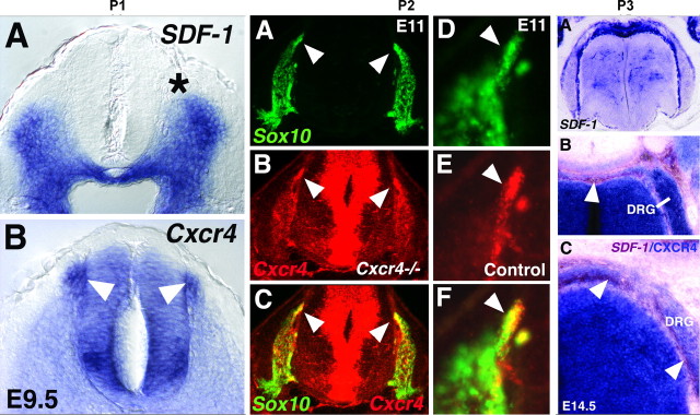
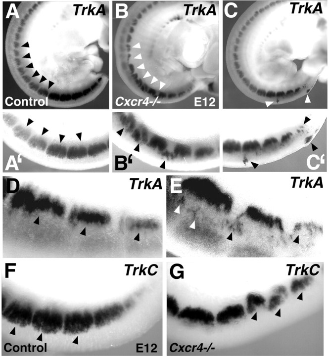
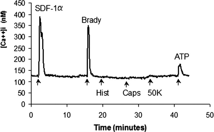
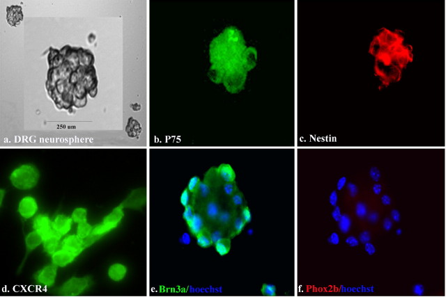
Chemokines and their receptors are essential for the development and organization of the hematopoietic/lymphopoietic system and have now been shown to be expressed by different types of cells in the nervous system. In mouse embryos, we observed expression of the chemokine (CXC motif) receptor 4 (CXCR4) by neural crest cells migrating from the dorsal neural tube and in the dorsal root ganglia (DRGs). Stromal cell-derived factor-1 (SDF-1), the unique agonist for CXCR4, was expressed along the path taken by crest cells to the DRGs, suggesting that SDF-1/CXCR4 signaling is needed for their migration. CXCR4 null mice exhibited small and malformed DRGs. Delayed migration to the DRGs was suggested by ectopic cells expressing tyrosine receptor kinase A (TrkA) and TrkC, neurotrophin receptors required by DRG sensory neuron development. In vitro, the CXCR4 chemokine receptor was upregulated by migratory progenitor cells just as they exited mouse neural tube explants, and SDF-1 acted as a chemoattractant for these cells. Most CXCR4-expressing progenitors differentiated to form sensory neurons with the properties of polymodal nociceptors. Furthermore, DRGs contained a population of progenitor cells that expressed CXCR4 receptors in vitro and differentiated into neurons with a similar phenotype. Our findings indicate an important role for SDF-1/CXCR4 signaling in directing the migration of sensory neuron progenitors to the DRG and potentially in other aspects of development once the DRGs have coalesced.
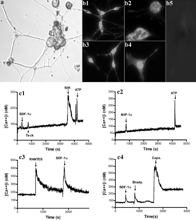

Figures

Figure 1.
Figure 1.
The expression patterns of SDF-1, CXCR4, and Sox10 within the spinal cords of mouse embryos. P1, A, B, Coronal sections through the wild-type mouse mid-spinal cord at E9.5, processed with in situ hybridization to show mRNA expression of SDF-1 and CXCR4. SDF-1 expression extends close to the site at which neural crest cells emerge from the neural tube (A, asterisk). The latter cells are marked by prominent expression of CXCR4 (B, arrowheads). Marked expression also appears in a distinct region of the ventral spinal cord, which may approximately correspond to the position of developing motor neurons. P2, A-F, Migrating DRG neural crest cells are CXCR4 positive. Sections through the spinal cord of a CXCR4 mutant (A-C) and a littermate control (D-F) at E11. Sections were processed for double-fluorescence in situ hybridization (Sox10, green, encodes a transcription factor expressed in neural crest cells; CXCR4, red). A-C, In a CXCR4 homozygous mutant, single fluorescence for Sox10 marks neural crest cells in and migrating to the DRGs on either side of the spinal cord (A). Arrowheads point to a patch of migrating cells also labeled for CXCR4 fluorescence (B, C). C is an overlay of A and B. D-F, Higher magnification of a Sox10- D, F; arrowhead) and CXCR4- (E, F) expressing cell group migrating toward the DRG (F is an overlay of D, E). P3, A-C, Coronal sections through wild-type E14.5 spinal cord processed with one- or two-color in situ hybridization for SDF-1 (blue in A) or both SDF-1 (brown in B, C) and CXCR4 (blue in B, C). SDF-1 expression is strong over the dorsal neural tube and runs ventrally in two streams that appear to surround the DRGs (B, C; arrowheads). Note that at this later age, E14.5, neural crest migration to the DRGs is near completion.
Figure 2.
Figure 2.
Abnormal DRG development in the absence of CXCR4 receptors. Whole-mount E12 embryos processed to show DRG expression of TrkA (A-C′, D, E) and TrkC (F, G). A-C′ are high-magnification views of the bottom third of A-C. In contrast with the regularly shaped DRGs forming in control embryos (A, A′), DRGs in CXCR4 null mutants are misshapen, disorganized, misshapen, or small (B, B′, C, C′). Arrowheads indicate normal DRGs (A, A′), compared with split, misshapen DRGs and cell islands in B, B′, C, and C′. Ectopic cells dorsal to the DRGs are indicated in E but are absent from the more regular DRGs in a control mouse (D). Similarly, DRGs marked by expression of TrkC are regularly shaped in a control mouse (F) but split or malformed in a CXCR4 null mouse (G).
Figure 3.
Figure 3.
Characterization of MNCs. A shows a trunk neural tube explant (Ne) at E9.5 with migrating cells exiting neural tube fragments. B, After replating, these cells formed nonadherent neurospheres. Scale bar, 150 μm. Also illustrated are immunostaining of migrating neural crest cells for p75 (C), nestin (D), CXCR4 (E, F), Brn3a (G), and Phox2b (H).
Figure 4.
Figure 4.
SDF-1 induces [Ca2+]i changes in MNCs. Trunk MNCs responded to SDF-1α, bradykinin (Brady), and ATP but not to capsaicin (Caps), high K [50 mm (50K)], or histamine (Hist). ATP-induced Ca responses were present in all cells tested. A total of 2340 cells of 3465 (67.5%) showed [Ca2+]i increase when stimulated by SDF-1α and 3255 of 3735 (87%) by bradykinin.
Figure 5.
Figure 5.
SDF-1 is a chemoattractant for migrating neural crest cells. a, Trunk MNCs migrated toward an SDF-1α (20 nm) gradient (90 min). Migration was inhibited by AMD3100 (AMD; 20 μm) (n = 4). Error bars represent SEM. *p < 0.05, significantly different from the control group; **p < 0.05, significantly different from the SDF-1-treated group. b, c, Neural crest cell migration from neural tube explant cultures. Neural crest cells rapidly emerged from explanted neural tube fragments and migrated out as a monolayer of cells surrounding the central dense mass of neuroepithelial tissue. Note the increase in the area of the neural crest outgrowth from 24 h (b) to 48 h (c) in culture. d, Insitu hybridization using SDF-1 mRNA probe showing expression of SDF-1 by a population of MNCs. e, Negative control for SDF-1 sense riboprobe.
Figure 6.
Figure 6.
Characteristics of differentiated trunk MNCs. a, Trunk MNCs were differentiated with NGF plus LIF (100 ng/ml) for 2 weeks. Immunostaining for CXCR4 (b), Brn3a (c), and Phox2b (d). Nuclear counterstaining with Hoechst 33342 (hoechst; blue) in b and d. e, Changes in [Ca2+]i in response to chemokines. Note that neurons exhibited responses to capsaicin (Caps) and to 50 mm K (50K; compare with Fig. 3). A total of 280 cells of 540 (52%) showed [Ca2+]i increase when stimulated by SDF-1α, 520 of 540 (96%) by bradykinin (Brady), and 380 of 540 (71%) by capsaicin.
Figure 7.
Figure 7.
Characterization of dividing DRG neurospheres. a, Phase-contrast illustration of a DRG neurosphere from a 7-d-old culture of an E14.5 mouse DRG. Anti-p75 staining (b), anti-nestin (c), anti-CXCR4 (d), anti-Brn3a with Hoechst 33342 (hoechst) counterstain in blue (e), and same neurosphere shown in e assessed for Phox2b staining with Hoescht 33342 counterstain in blue (f).
Figure 8.
Figure 8.
DRG neurospheres express a variety of chemokine receptors. A, RT-PCR products indicating that different chemokine receptors are expressed by E14.5 DRG neurospheres. pb, Pair base. B, [Ca2+]i changes in response to chemokines in dividing DRG neurospheres. A total of 170 cells of 540 (32%) showed [Ca2+]i increases in response to SDF-1α. Note that there were no responses to either 50 mm K (50K) or to capsaicin (Caps). Rantes, Regulated upon Activation, Normal T cell Expressed and Secreted; Teck, thymus expressed chemokine; Fract, Fractalkine; Ctack, cutaneous T cell-attracting chemokine; Hist, histamine.
Figure 9.
Figure 9.
Characteristics of differentiated DRG neurospheres. a, Differentiated DRG neurosphere cells after culture with NGF and LIF (100 ng/ml) for 7-10 d. Immunostaining of differentiated DRG cells for CXCR4 (b1, b2), Brn3a (b3, b4), and Phox2b (b5). DRG cells gave rise to both neuronal cells (b1, b3) and flat cells (b2, b4) that were positive for CXCR4 and Brn3a. b5 indicates lack of staining for Phox2b. Changes in [Ca2+]i in response to 50 mm K (50K), chemokines, and other agonists in differentiated DRG cells (c1-c4). A total of 130 cells of 420 (31%) showed [Ca2+]i increase when stimulated by SDF-1α, 110 of 160 (69%) by bradykinin (Brady.), and 260 of 400 (65%) by capsaicin (Caps.).

References

    1. Agarwala S, Sanders TA, Ragsdale CW (2001) Sonic hedgehog control of size and shape in midbrain pattern formation. Science 291: 2147-2150. - PubMed
    1. An M, Luo R, Henion PD (2002) Differentiation and maturation of zebrafish dorsal root and sympathetic ganglion neurons. J Comp Neurol 446: 267-275. - PubMed
    1. Anderson DJ (2000) Genes, lineages and the neural crest: a speculative review. Philos Trans R Soc Lond B Biol Sci 355: 953-964. - PMC - PubMed
    1. Bagri A, Gurney T, He X, Zou YR, Littman DR, Tessier-Lavigne M, Pleasure SJ (2002) The chemokine SDF1 regulates migration of dentate granule cells. Development 129: 4249-4260. - PubMed
    1. Bodner A, Toth PT, Miller RJ (2004) Activation of c-Jun N-terminal kinase mediates gp120IIIB- and nucleoside analogue-induced sensory neuron toxicity. Exp Neurol 118: 246-253. - PubMed

Publication types

MeSH terms

Substances

LinkOut - more resources