Comparative Study
. 2005 Apr 20;25(16):4031-9.
doi: 10.1523/JNEUROSCI.4969-04.2005.

Presynaptically silent GABA synapses in hippocampus

Affiliations
Comparative Study

Presynaptically silent GABA synapses in hippocampus

John M Bekkers. J Neurosci. .

Abstract

Mammalian central synapses commonly specialize in one fast neurotransmitter, matching the content of their presynaptic vesicles with the appropriate receptors in their postsynaptic membrane. Here, I show that hippocampal cultures contain autaptic glutamatergic synapses that contravene this rule: in addition to postsynaptic glutamate receptors, they also express clusters of functional postsynaptic GABA(A) receptors yet lack presynaptic GABA. Hence, these synapses are presynaptically silent with respect to GABA. They can be unsilenced by loading GABA into presynaptic vesicles by endocytosis, after which a postload IPSC appears. This IPSC is similar to native IPSCs recorded from GABAergic interneurons in the same cultures. Thus, these "mistargeted" GABA(A) receptors, which apparently lack a signal that confers synaptic specificity, function almost normally. After GABA loading, glutamatergic miniature postsynaptic currents acquire a slow tail that is mediated by GABA(A) receptors, showing that synaptic vesicles can accommodate both the usual concentration of native glutamate and a saturating concentration of loaded GABA. After brief Ca(2+)-dependent exocytosis, endocytosis of GABA can proceed in low-Ca(2+) external solution. The amplitude of the postload IPSC declines exponentially with repetitive stimulation as the endocytosed GABA passes through the presynaptic vesicle cycle and is depleted. Hence, by using GABA as an exogenous but physiological tracer, the properties of these presynaptically silent synapses can provide novel insights into the content and cycling of vesicles in presynaptic terminals.


Figures
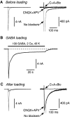
Figure 1.
Figure 1.
A postload IPSC appears after GABA loading. A, Before loading, the autaptic EPSC (large gray trace; No blockers) is almost fully blocked by 20 μm CNQX plus 50 μm d-APV (black trace). The addition of 20 μm bicuculline (flat gray trace; C+A+Bic) has little additional effect. The two panels show the same traces on different vertical scales. B, The cell was superfused for 90 s with a loading solution (in mm: 100 GABA, 2 CaCl2, and 40 KCl) that produced a large, desensitizing whole-cell GABA current. (The current is inward because the electrode solution contains a high chloride concentration.) C, After washing out the loading solution and resuming autaptic stimulation, the original EPSC reappears. Now, addition of CNQX plus d-APV reveals a large postload IPSC (black trace) that is blocked by bicuculline.
Figure 3.
Figure 3.
Postload IPSCs behave presynaptically like glutamatergic synapses. A, Plot of native EPSC amplitude (Amp; measured at 2 s intervals) versus time. Bath-applied adenosine (100 μm) reversibly inhibits the EPSC. The right panel shows example traces at the numbered time points. B, Plot of native IPSC amplitude (measured at 10 s intervals) versus time. Adenosine (100 μm) has no effect. C, Plot of postload IPSC amplitude (2 s intervals; same cell as in A) versus time. Adenosine (100 μm) reversibly inhibits the postload IPSC. Bic, Bicuculline. D, Summary of Ado experiments. Both native EPSCs and postload IPSCs are significantly inhibited (*p < 0.05) by 100 μm adenosine, unlike native IPSCs. Error bars represent SEM.
Figure 5.
Figure 5.
GABA can be endocytosed in the absence of Ca2+, provided Ca2+-dependent exocytosis has occurred previously. In this figure, the black traces are bicuculline-sensitive autaptic currents recorded in CNQX plus d-APV, and the gray traces are whole-cell currents elicited by bath application of the indicated solutions. A, After superfusion of a high-K+ GABA loading solution that lacks added Ca2+, there is little change in the autaptic current (middle black trace). Next, the same cell was superfused briefly (5 s) with a solution containing 2 mm Ca2+ and 40 mm K+ but no GABA to trigger exocytosis. The bath solution was then immediately switched to one containing 100 mm GABA and normal K+ but no added Ca2+. After endocytosis for 60 s in this Ca2+-free loading solution, a large postload IPSC emerged (right black trace). B, Exocytosis was triggered by a train of 300 autaptic stimuli at 20 Hz in normal Ca2+-containing bath solution. The inset (gray trace) shows the beginning and end of this train. Starting 1.5 s after the end of the train, the cell was superfused for 60 s with nominally Ca2+-free GABA loading solution. After this, a large postload IPSC emerged (right black trace).
Figure 6.
Figure 6.
The amplitude of the postload IPSC irreversibly runs down with time, reflecting the passage of loaded GABA through the synaptic vesicle cycle. A, Plot of amplitude versus time for a typical experiment. The native EPSC amplitude was initially ∼25 nA [points near “1;” stimulus (Stim) rate 0.1 Hz]. After loading (40 mm K+, 100 mm GABA for 30 s), the cell was bathed in normal external solution containing CNQX plus d-APV to isolate the postload IPSC and then stimulated at 1 Hz (0-10 min). The amplitude of the IPSC declined exponentially [superimposed fit of a single exponential plus an offset; τ = 152 s; baseline confirmed by addition of bicuculline (+Bic)]. The stimulus rate was then reduced to 0.1 Hz (10-15 min) to confirm that the reduction in IPSC amplitude was irreversible and not simply caused by temporary exhaustion of the readily releasable pool of synaptic vesicles at the higher stimulation rate. Finally, the CNQX plus d-APV was washed out, confirming that the native EPSC was unchanged in amplitude (points near “2”). B, Single synaptic currents recorded near the numbered time points in A.
Figure 4.
Figure 4.
Presynaptic vesicles at glutamatergic autapses can accommodate both loaded GABA and the normal concentration of glutamate. A, Individual miniature synaptic currents recorded in an isolated pyramidal neuron before GABA loading. The right panel shows normalized averaged minis recorded before (black; n = 42 minis) and during (gray, obscured; n = 56) application of 20 μm bicuculline. B, After loading, minis with a slow component (arrows) appear. The averages (right) show that the slow component is blocked by 20 μm bicuculline (Bic) (n = 48 minis before bicuculline; n = 41 after bicuculline). C, Dose-response plot of the amplitude of the evoked postload IPSC versus the concentration of GABA in the high-K+ loading solution. For each cell, the postload IPSC amplitude was expressed as a percentage of the native EPSC amplitude measured in that cell. This was done to normalize to the number of autapses on different cells. Each point is an average from five to seven cells. The superimposed logistic equation gives a half-maximal GABA concentration of 48 mm. Error bars represent SEM. D, Amplitude (Amp) histograms of minis recorded in bicuculline in the same cell before and after loading with 100 mm GABA. The amplitude distributions are not significantly different (p = 0.45; Kolmogorov-Smirnov test), suggesting that the presence of GABA in some vesicles does not affect the amount of glutamate that can be accommodated.
Figure 2.
Figure 2.
Normal perisynaptic GABAA receptors mediate the postload IPSC. A, Examples of a native autaptic IPSC recorded in an isolated GABAergic interneuron (gray trace) and a postload IPSC recorded in an isolated pyramidal neuron (black trace) scaled so their peak amplitudes are equal. The rise time of the postload IPSC is slower than that of the native IPSC (left), but the decay is identical (right). B, Summary of kinetic measurements. The averaged 10-90% rise time (RT) for postload IPSCs (bar 2, left) is significantly greater than that for native IPSCs (bar 1; *p < 0.05). This is not caused by a systematic voltage-clamp error between interneurons and pyramidal neurons, because the rise time for native EPSCs (bar 3), measured in the same cells as the postload IPSCs, is as fast as that for native IPSCs. C, Pentobarbital (20 μm) reversibly slows the decay of both native (left traces) and postload (right traces) IPSCs. c, Control; PB, pentobarbital; w, washout. D, Summary of PB experiments. The weighted decay time constant of native and postload IPSCs is equally prolonged by PB. Error bars represent SEM.

Similar articles

Cited by

References

    1. Aravanis AM, Pyle JL, Tsien RW (2003) Single synaptic vesicles fusing transiently and successively without loss of identity. Nature 423: 643-647. - PubMed
    1. Axmacher N, Stemmler M, Engel D, Draguhn A, Ritz R (2004) Transmitter metabolism as a mechanism of synaptic plasticity: a modeling study. J Neurophysiol 91: 25-39. - PubMed
    1. Banks MI, Pearce RA (2000) Kinetic differences between synaptic and extrasynaptic GABAA receptors in CA1 pyramidal cells. J Neurosci 20: 937-948. - PMC - PubMed
    1. Bekkers JM, Stevens CF (1991) Excitatory and inhibitory autaptic currents in isolated hippocampal neurons maintained in cell culture. Proc Natl Acad Sci USA 88: 7834-7838. - PMC - PubMed
    1. Betz WJ, Mao F, Bewick GS (1992) Activity-dependent fluorescent staining and destaining of living vertebrate motor nerve terminals. J Neurosci 12: 363-375. - PMC - PubMed

Publication types

MeSH terms

LinkOut - more resources