Comparative Study
. 2005 Apr 20;25(16):4082-90.
doi: 10.1523/JNEUROSCI.4306-04.2005.

Thrombin-induced oxidative stress contributes to the death of hippocampal neurons in vivo: role of microglial NADPH oxidase

Affiliations
Comparative Study

Thrombin-induced oxidative stress contributes to the death of hippocampal neurons in vivo: role of microglial NADPH oxidase

Sang-Ho Choi et al. J Neurosci. .

Abstract

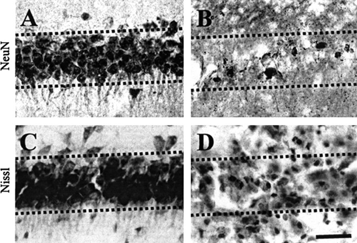
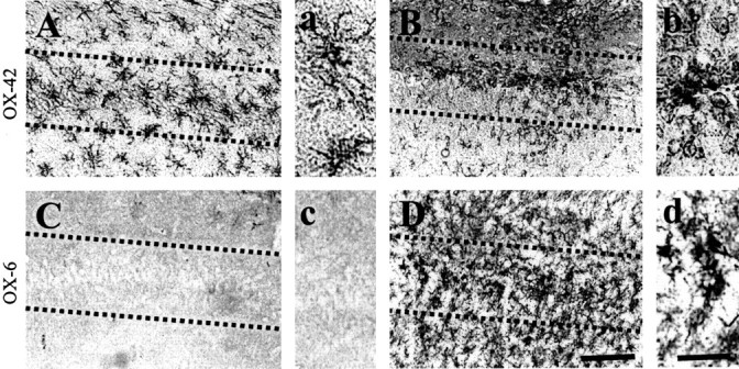
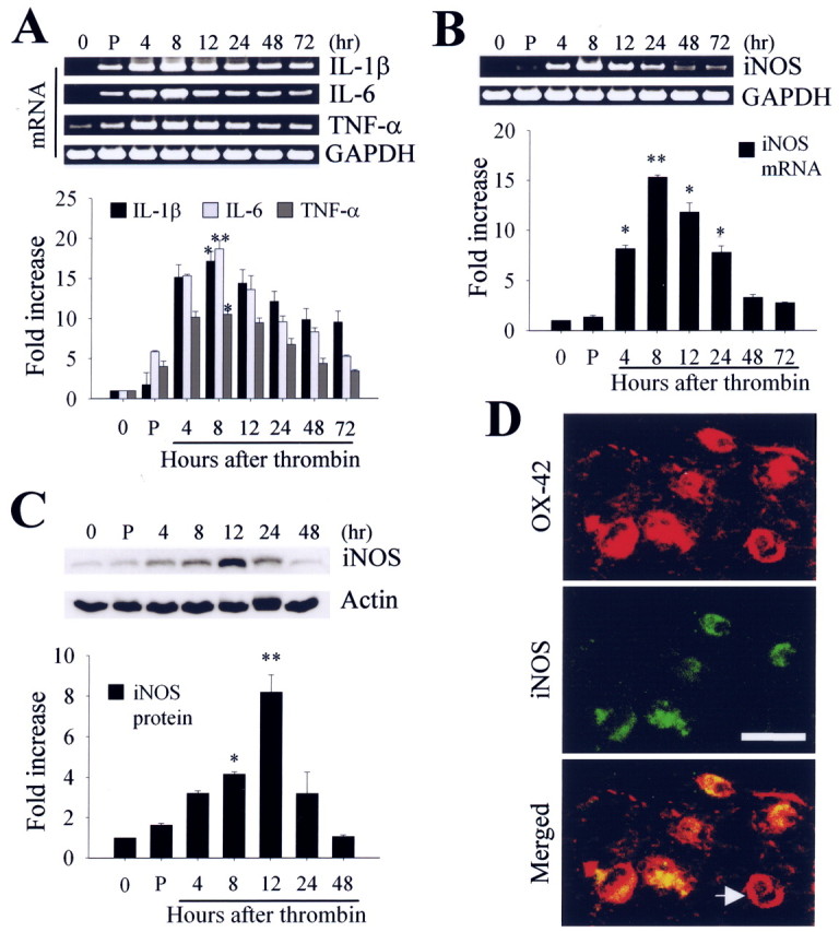
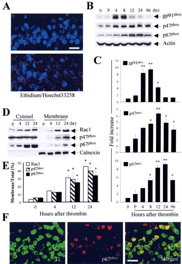
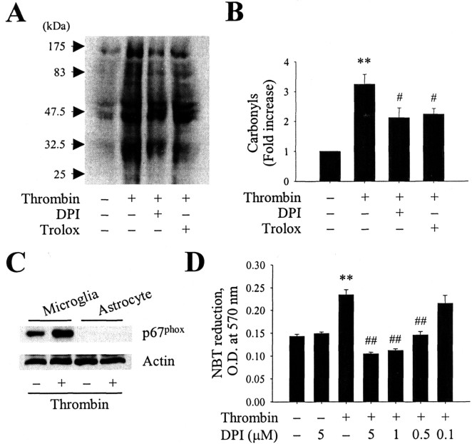
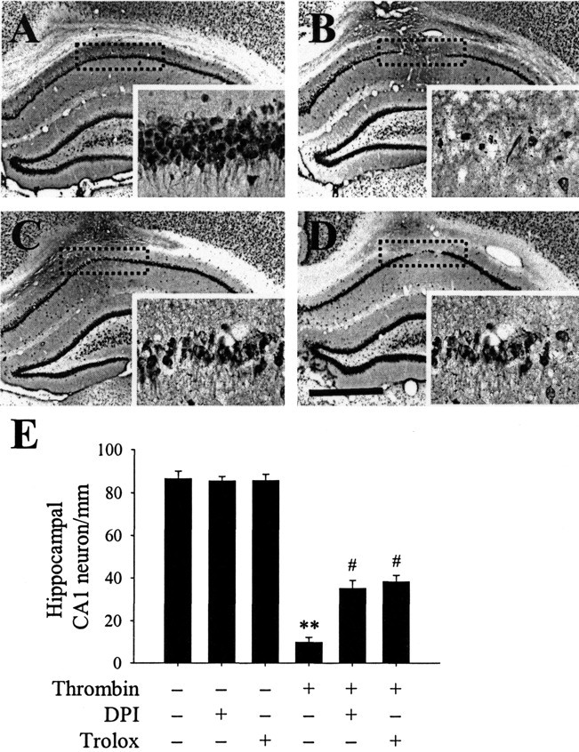
The present study investigated whether thrombin, a potent microglial activator, can induce reactive oxygen species (ROS) generation through activation of microglial NADPH oxidase and if this may contribute to oxidative damage and consequent neurodegeneration. Seven days after intrahippocampal injection of thrombin, Nissl staining and immunohistochemistry using the neuronal-specific nuclear protein NeuN revealed a significant loss in hippocampal CA1 neurons. In parallel, thrombin-activated microglia, assessed by OX-42 and OX-6 immunohistochemistry, and ROS production, assessed by hydroethidine histochemistry, were observed in the hippocampal CA1 area in which degeneration of hippocampal neurons occurred. Reverse transcription-PCR at various time points after thrombin administration demonstrated an early and transient expression of inducible nitric oxide synthase (iNOS) and several proinflammatory cytokines. Western blot analysis and double-label immunohistochemistry showed an increase in the expression of and the localization of iNOS within microglia. Additional studies demonstrated that thrombin induced the upregulation of membrane (gp91(phox)) and cytosolic (p47(phox) and p67(phox)) components, translocation of cytosolic proteins (p47(phox), p67(phox), and Rac1) to the membrane, and p67(phox) expression of the NADPH oxidase in microglia in the hippocampus in vivo, indicating the activation of NADPH oxidase. The thrombin-induced oxidation of proteins and loss of hippocampal CA1 neurons were partially inhibited by an NADPH oxidase inhibitor and by an antioxidant. To our knowledge, the present study is the first to demonstrate that thrombin-induced neurotoxicity in the hippocampus in vivo is caused by microglial NADPH oxidase-mediated oxidative stress. This suggests that thrombin inhibition or enhancing antioxidants may be beneficial for the treatment of neurodegenerative diseases, such as Alzheimer's disease, that are associated with microglial-derived oxidative damage.


Figures

Figure 1.
Figure 1.
Thrombin-induced neurotoxicity in the CA1 layer of rat hippocampus in vivo. PBS (A, C) or thrombin (B, D; 20 U/4 μl) was unilaterally injected into the CA1 layer of the hippocampus. Animals were killed 7 d after injection, brains were removed, and coronal sections (40 μm) were cut using a sliding microtome. Every sixth serial section was selected and processed for NeuN immunostaining or Nissl staining. A, B, NeuN immunostaining in the CA1 layer of hippocampus. Note that there is a significant reduction of NeuN-immunopositive cells in thrombin-injected hippocampus. C, D, CA1 layer of the hippocampus stained for Nissl substance (cresyl violet). The results are representative of six to eight animals per group. Dotted lines indicate the CA1 layer of the hippocampus. Scale bar, 200 μm.
Figure 2.
Figure 2.
Thrombin-induced microglial activation in the CA1 area of the hippocampus. PBS (A, C) or thrombin (B, D; 20 U/4 μl) was unilaterally injected into the CA1 layer of the hippocampus. Animals were killed 24 h after injection, brains were removed, and coronal sections were cut using a sliding microtome. Brain sections were immunostained with OX-42 (A, B) or OX-6 (C, D) antibodies to identify microglia. a-d show higher magnifications of A-D, respectively. Note the significant microglial activation in the thrombin-treated CA1 area of the hippocampus compared with the PBS-treated control (A, C). These results are representative of six to eight animals per group. Dotted lines indicate the CA1 cell layer of the hippocampus. Scale bars: A-D, 200 μm; a-d, 50 μm.
Figure 3.
Figure 3.
A, B, Reverse transcription-PCR analysis of thrombin-induced mRNA expression of proinflammatory cytokines and iNOS in the hippocampus. Animals were decapitated after intrahippocampal injection of thrombin (20 U/4 μl), and total RNA was isolated in the ipsilateral hippocampus at the indicated time points. C, Western blot analysis of iNOS expression in hippocampus at indicated time points after intrahippocampal thrombin injection. In A-C, untreated (0 h) or PBS-treated (4 h) hippocampus was used as a control. Error bars represent the mean ± SEM for four to five samples per time point. *p < 0.05, **p < 0.01 compared with control according to ANOVA and Student-Newman-Keuls analyses. P, PBS. D, Colocalization of iNOS immunoreactivity within the activated microglia in the CA1 area of hippocampus. The sections of rat hippocampus were prepared 12 h after intrahippocampal injection of thrombin (20 U/4 μl) and then immunostained simultaneously with iNOS and OX-42 as a marker of microglia. Images were captured from the same area and merged. Note the absence of iNOS expression in some microglia (indicated by an arrow). Scale bar, 25 μm.
Figure 4.
Figure 4.
A, In situ visualization of thrombin-induced O2- and O2--derived oxidant production in the CA1 layer of the hippocampus. Animals were injected with hydroethidine (1 μg/μl, i.p.) 48 h after intrahippocampal injection of thrombin (20 U/4 μl). After 15 min, the animals were killed, and sections of hippocampus were prepared for hydroethidine histochemistry to detect the extracellular superoxide. Confocal micrographs show ethidium fluorescence (red) in PBS-injected (top) or thrombin-injected (bottom) CA1 region of the hippocampus. Nuclei were counterstained with Hoechst 33258 (blue). B, Western blot analysis showing upregulation of membrane-bound subunit gp91phox and cytosolic subunits p47phox and p67phox in thrombin-treated hippocampus. Animals were decapitated after intrahippocampal injection of thrombin (20 U) at the indicated time points, and the hippocampi were isolated immediately from the ipsilateral hemisphere. Untreated (0 h) or PBS-treated (4 h) hippocampus was used as a control. Tissue lysates were analyzed by Western blot analysis with gp91phox, p47phox, and p67phox antibodies. C, Error bars represent the mean ± SEM from four to five samples per time point. *p < 0.05, **p < 0.01 compared with control according to ANOVA and Student-Newman-Keuls analyses. D, Translocation of cytosolic subunits (Rac1, p47phox, and p67phox) from the cytosol to the plasma membrane after intrahippocampal injection of thrombin, indicating activation of NADPH oxidase in the hippocampus. Animals were decapitated after intrahippocampal injection of thrombin (20 U) at the indicated time points, and hippocampi were isolated immediately from the ipsilateral hemisphere. Tissue lysates were fractionated and analyzed by immunoblot analysis with p67phox antibody. The membrane protein calnexin was used to normalize the data. E, Error bars represent the mean ± SEM for four to five samples per time point. The levels of NADPH oxidase subunits in the cytosol and membrane were determined by densitometric scanning of Western blots, and the levels of those subunits in the membrane were expressed as a percentage of the total level of each subunit. *p < 0.05, **p < 0.01 compared with control according to ANOVA and Student-Newman-Keuls analyses. F, Localization of p67phox immunoreactivity in activated microglia within the CA1 region of the hippocampus treated with thrombin. The sections of hippocampus were prepared 12 h after thrombin injection (20 U) and then simultaneously stained with an antibody against p67phox and a maker for microglia (tomato lectin). Confocal images were captured from the same area and merged. TL, Tomato lectin. Scale bars: A, 100 μm; F, 50 μm.
Figure 5.
Figure 5.
A, Thrombin induces protein oxidation in the hippocampus. Animals were decapitated 48 h after intrahippocampal injection of thrombin (20 U) in the absence or presence of the NADPH oxidase inhibitor DPI (100 μm, i.c.v.) or the antioxidant trolox (50 mg/kg, i.p.). The hippocampi were isolated immediately from the ipsilateral hemisphere. Samples were analyzed by Western blotting for protein carbonyls as markers of oxidatively modified proteins. B, Bars represent the means ± SEM of four to five samples. *p < 0.05, **p < 0.01 compared with control according to ANOVA and Student-Newman-Keuls analyses. C, Western blot analysis showing expression of p67phox protein in rat cortical cultures of microglia or astrocytes treated for 6 h in the absence or presence of thrombin (40 U/ml). D, Effect of DPI on thrombin-induced production of O2- in cultured microglia. Cultures of microglia were pretreated for 1 h with DPI (0.1-5 μm), after which they were treated for 12 h with thrombin (40 U/ml). Next, the cells were treated for 1 h with nitroblue tetrazolium (NBT; 1 mg/ml), the medium was removed, and the resulting formazan was dissolved in dimethylsulfoxide. The lysates were transferred to a 96-well plate, and the absorbance was measured at 570 nm in a spectrophotometer. Error bars represent the mean ± SEM of triplicate cultures in the three separate platings. **p < 0.01 compared with untreated control cultures; ##p < 0.01 compared with cultures treated with thrombin only (ANOVA and Student-Newman-Keuls analyses).
Figure 6.
Figure 6.
DPI or trolox prevents thrombin-induced neuronal death in the hippocampus. PBS or thrombin (20 U) was unilaterally injected into the hippocampus in either the absence or presence of the NADPH oxidase inhibitor DPI (100 μm, i.c.v.) or the antioxidant trolox (50 mg/kg, i.p.). Animals were killed 7 d after injection. The brains were removed, and coronal sections (40 μm) were cut using a sliding microtome. Every sixth serial section was selected and processed for NeuN immunostaining. Shown are images of a hippocampus treated with PBS alone (A), thrombin (B), DPI plus thrombin (C), or trolox plus thrombin (D). Insets show magnified photomicrographs of the area in the CA1 layer marked by dotted rectangles. The results are representative of six to eight animals per group. Scale bar, 1 mm. E, Number of NeuN-immunopositive neurons in the CA1 layer of hippocampi treated with thrombin in either the absence or presence of DPI or trolox. As controls, DPI or trolox alone was injected. Error bars represent the means ± SEM from six to eight samples per group. **p < 0.01 compared with untreated hippocampi; #p < 0.05 compared with hippocampi treated with thrombin only (ANOVA and Student-Newman-Keuls analyses).

Similar articles

Cited by

References

    1. Abramov AY, Canevari L, Duchen MR (2004) β-Amyloid peptides induce mitochondrial dysfunction and oxidative stress in astrocytes and death of neurons through activation of NADPH oxidase. J Neurosci 24: 565-575. - PMC - PubMed
    1. Akiyama H, Ikeda K, Kondo H, McGeer PL (1992) Thrombin accumulation in brains of patients with Alzheimer's disease. Neurosci Lett 146: 152-154. - PubMed
    1. Akiyama H, Barger S, Barnum S, Bradt B, Bauer J, Cole GM, Cooper NR, Eikelenboom P, Emmerling M, Fiebich BL, Finch CE, Frautschy S, Griffin WS, Hampel H, Hull M, Landreth G, Lue L, Mrak R, Mackenzie IR, McGeer PL, et al. (2000) Inflammation and Alzheimer's disease. Neurobiol Aging 21: 383-421. - PMC - PubMed
    1. Babior BM (1999) NADPH oxidase: an update. Blood 93: 1464-1476. - PubMed
    1. Bal-Price A, Matthias A, Browin GC (2002) Stimulation of NADPH oxidase in activated rat microglia removes nitric oxide but induces peroxynitrite production. J Neurochem 80: 73-80. - PubMed

Publication types

MeSH terms