

Stereophysicochemical variability plots highlight conserved antigenic areas in Flaviviruses
- PMID: 15845145
- PMCID: PMC1112618
- DOI: 10.1186/1743-422X-2-40
Stereophysicochemical variability plots highlight conserved antigenic areas in Flaviviruses
Abstract
Background: Flaviviruses, which include Dengue (DV) and West Nile (WN), mutate in response to immune system pressure. Identifying escape mutants, variant progeny that replicate in the presence of neutralizing antibodies, is a common way to identify functionally important residues of viral proteins. However, the mutations typically occur at variable positions on the viral surface that are not essential for viral replication. Methods are needed to determine the true targets of the neutralizing antibodies.
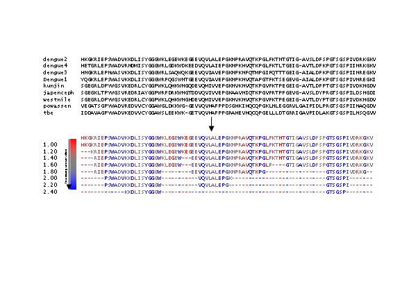
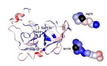
Results: Stereophysicochemical variability plots (SVPs), 3-D images of protein structures colored according to variability, as determined by our PCPMer program, were used to visualize residues conserved in their physical chemical properties (PCPs) near escape mutant positions. The analysis showed 1) that escape mutations in the flavivirus envelope protein are variable residues by our criteria and 2) two escape mutants found at the same position in many flaviviruses sit above clusters of conserved residues from different regions of the linear sequence. Conservation patterns in T-cell epitopes in the NS3- protease suggest a similar mechanism of immune system evasion.
Conclusion: The SVPs add another dimension to structurally defining the binding sites of neutralizing antibodies. They provide a useful aid for determining antigenically important regions and designing vaccines.
Figures
Similar articles
-
Structural basis of a flavivirus recognized by its neutralizing antibody: solution structure of the domain III of the Japanese encephalitis virus envelope protein.J Biol Chem. 2003 Nov 14;278(46):46007-13. doi: 10.1074/jbc.M307776200. Epub 2003 Sep 2. J Biol Chem. 2003. PMID: 12952958
-
Second site escape of a T20-dependent HIV-1 variant by a single amino acid change in the CD4 binding region of the envelope glycoprotein.Retrovirology. 2006 Nov 29;3:84. doi: 10.1186/1742-4690-3-84. Retrovirology. 2006. PMID: 17134507 Free PMC article.
-
Characterization of neutralizing sites in the second variable and fourth variable region in gp125 and a conserved region in gp36 of human immunodeficiency virus type 2.Viral Immunol. 1999;12(1):79-88. doi: 10.1089/vim.1999.12.79. Viral Immunol. 1999. PMID: 10333245
-
Antigenic structure of flavivirus proteins.Adv Virus Res. 2003;59:141-75. doi: 10.1016/s0065-3527(03)59005-4. Adv Virus Res. 2003. PMID: 14696329 Review.
-
Domain III peptides from flavivirus envelope protein are useful antigens for serologic diagnosis and targets for immunization.Biologicals. 2010 Nov;38(6):613-8. doi: 10.1016/j.biologicals.2010.07.004. Biologicals. 2010. PMID: 20817489 Review.
Cited by
-
Base of the measles virus fusion trimer head receives the signal that triggers membrane fusion.J Biol Chem. 2012 Sep 21;287(39):33026-35. doi: 10.1074/jbc.M112.373308. Epub 2012 Aug 2. J Biol Chem. 2012. PMID: 22859308 Free PMC article.
-
Identification and analysis of conserved sequence motifs in cytochrome P450 family 2. Functional and structural role of a motif 187RFDYKD192 in CYP2B enzymes.J Biol Chem. 2008 Aug 1;283(31):21808-16. doi: 10.1074/jbc.M708582200. Epub 2008 May 21. J Biol Chem. 2008. PMID: 18495666 Free PMC article.
-
PCP consensus sequences of flaviviruses: correlating variance with vector competence and disease phenotype.J Mol Biol. 2010 Feb 26;396(3):550-63. doi: 10.1016/j.jmb.2009.11.070. Epub 2009 Dec 4. J Mol Biol. 2010. PMID: 19969003 Free PMC article.
-
Small molecule grp94 inhibitors block dengue and Zika virus replication.Antiviral Res. 2019 Nov;171:104590. doi: 10.1016/j.antiviral.2019.104590. Epub 2019 Aug 14. Antiviral Res. 2019. PMID: 31421166 Free PMC article.
-
AllerTOP v.2--a server for in silico prediction of allergens.J Mol Model. 2014 Jun;20(6):2278. doi: 10.1007/s00894-014-2278-5. Epub 2014 May 31. J Mol Model. 2014. PMID: 24878803
References
-
- Holmes EC, Twiddy SS. The origin, emergence and evolutionary genetics of dengue virus. Infect Genet Evol. 2003;3:19–28. - PubMed
-
- Halstead SB, Deen J. The future of dengue vaccines. LANCET. 2002;360:1243–1245. - PubMed
-
- Mongkolsapaya J, Dejnirattisai W, Xu X, Vasanawathana S, Tangthawornchaikul N, Chairunsri A, Sawasdivorn S, Duangchinda T, Dong T, Rowland-Jones S, Yenchitsomanus P, McMichael A, Malasit P, Screaton G. Original antigenic sin and apoptosis in the pathogenesis of dengue hemorrhagic fever. Nature Med. 2003;9:921–927. - PubMed
Publication types
MeSH terms
Substances
Grants and funding
LinkOut - more resources
Full Text Sources
Other Literature Sources
