. 2005 May;73(5):2728-35.
doi: 10.1128/IAI.73.5.2728-2735.2005.

Outer membrane protein P6 of nontypeable Haemophilus influenzae is a potent and selective inducer of human macrophage proinflammatory cytokines

Affiliations

Outer membrane protein P6 of nontypeable Haemophilus influenzae is a potent and selective inducer of human macrophage proinflammatory cytokines

Charles S Berenson et al. Infect Immun. 2005 May.

Abstract

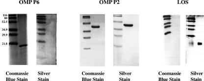
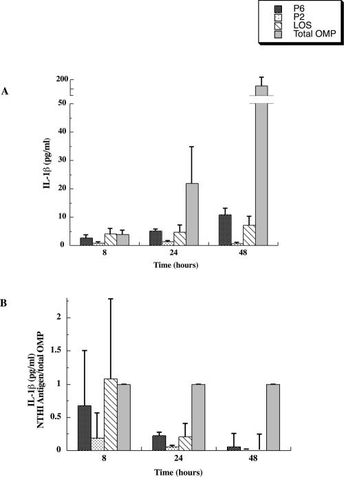
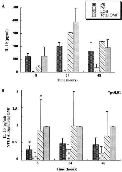
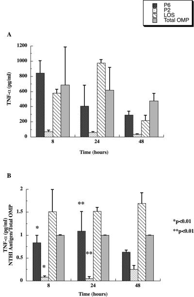
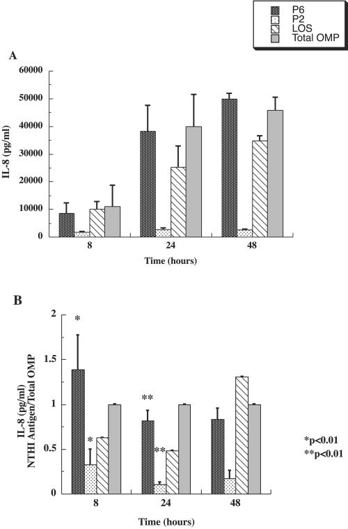
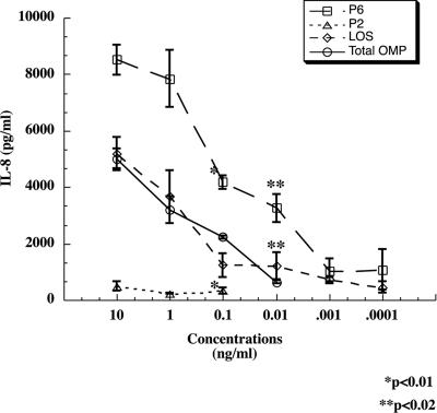
Interactions of nontypeable Haemophilus influenzae (NTHI) with human macrophages contribute to the pathogenesis of NTHI-induced infection in humans. However, the immunologic mechanisms that initiate and perpetuate NTHI-mediated macrophage responses have not been well explored. Outer membrane protein (OMP) P6 is a conserved lipoprotein expressed by NTHI in vivo that possesses a Pam(3)Cys terminal motif, characteristic of immunoactive bacterial lipoproteins associated with Toll-like receptor signaling. We theorized that OMP P6 is a potent immunomodulator of human macrophages. To test this hypothesis, we purified OMP P6 as well as OMP P2, the predominant NTHI outer membrane protein, and lipooligosaccharide (LOS), the specific endotoxin of NTHI, from NTHI strain 1479. Human blood monocyte-derived macrophages, purified from healthy donors, were incubated with each outer membrane constituent, and cytokine production of macrophage supernatants interleukin-1beta (IL-1beta), tumor necrosis factor alpha (TNF-alpha), IL-10, IL-12, and IL-8 was measured. OMP P6 selectively upregulated IL-10, TNF-alpha, and IL-8. While OMP P6 (0.1 mug/ml for 8 h) elicited slightly greater concentrations of IL-10, it resulted in over ninefold greater concentrations of TNF-alpha and over fourfold greater concentrations of IL-8 than did OMP P2. OMP P6 at doses as low as 10 pg/ml was still effective at induction of macrophage IL-8, while OMP P2 and LOS were not. OMP P6 of NTHI is a specific trigger of bacteria-induced human macrophage inflammatory events, with IL-8 and TNF-alpha as key effectors of P6-induced macrophage responses.


Figures

FIG. 1.
FIG. 1.
Purified preparations of NTHI outer membrane constituents. Purified preparations were made of OMP P6, OMP P2, and LOS of NTHI 1479. Purity of each a preparation (5 to 10 μg) was confirmed with Coomassie blue stain and by absence of other bands with silver stain in addition to Limulus assay for endotoxin. Molecular weight standards are shown to the left of each lane and are labeled in the first lane.
FIG. 2.
FIG. 2.
NTHI outer membrane antigen induction of macrophage IL-1β. Macrophages were incubated with OMP P6, OMP P2, LOS, or the total OMP preparation, each at 0.1 μg/ml, and IL-1β induction was measured for 8, 24, and 48 h. Results shown in panel A are means ± SEM from triplicate samples of one donor and were reproducible with cells from three separate donors. Combined results of all three donors, shown in panel B, are expressed as IL-1β concentration elicited with each antigen relative to IL-1β elicited by total OMPs. Macrophage reactivity was confirmed by treatment with E. coli K235LPS (1 μg/ml) for each time point.
FIG. 3.
FIG. 3.
NTHI outer membrane antigen induction of macrophage IL-12. Macrophages were incubated with each outer membrane constituent as in Fig. 2, and IL-12 induction was measured. Concentrations of each antigen and incubation times are as detailed in Fig. 2. Results shown in panel A are means ± SEM from triplicate samples of one donor. Combined results of all three donors, shown in panel B, are expressed as IL-12 concentration elicited with each antigen relative to IL-12 elicited by total OMPs.
FIG. 4.
FIG. 4.
NTHI outer membrane antigen induction of macrophage IL-10. Macrophages were incubated with each outer membrane constituent as in Fig. 2, and IL-10 induction was measured. Concentrations of each antigen and incubation times are as detailed in Fig. 2. Panel A shows means ± SEM of IL-10 from triplicate samples of one donor and were reproducible with cells from three separate donors. Panel B shows IL-10 concentration elicited with each antigen relative to IL-10 elicited by total OMPs to accommodate donor variability.
FIG. 5.
FIG. 5.
NTHI outer membrane antigen induction of macrophage TNF-α. Macrophages were incubated with each outer membrane constituents as in Fig. 2, and TNF-α induction was measured. Concentrations of each antigen and incubation times are as detailed in Fig. 2. Panel A shows means ± SEM of TNF-α from triplicate samples of one donor and were reproducible with cells from three separate donors. Results from all three donors are expressed in panel B as TNF-α concentration from each antigen relative to TNF-α concentration from the total OMP preparation.
FIG. 6.
FIG. 6.
NTHI outer membrane antigen induction of macrophage IL-8. Macrophages were incubated with each outer membrane constituent as in Fig. 2, and IL-8 induction was measured. Concentrations of each antigen and incubation times are as detailed in Fig. 2. Panel A shows means ± SEM of TNF-α from triplicate samples of one donor and were reproducible with cells from three separate donors. As with TNF-α measurements, relative differences between individual donors are accommodated by expressing values as IL-8 concentration elicited by each antigen relative to IL-8 concentration elicited by total OMPs in panel B.
FIG. 7.
FIG. 7.
Macrophage IL-8 induction with diminished concentrations of NTHI outer membrane antigens. Macrophages were incubated with decreasing concentrations of each outer membrane constituent. As in previous experiments, each value represents means ± SEM of three separate measurements.

Similar articles

Cited by

References

    1. Abe, Y., T. F. Murphy, S. Sethi, H. S. Faden, J. Dmochowski, Y. Harabuchi, and Y. M. Thanavala. 2002. Lymphocyte proliferative response to P6 of Haemophilus influenzae is associated with relative protection from exacerbations of chronic obstructive pulmonary disease. Am. J. Respir. Crit. Care Med. 165:967-971. - PubMed
    1. Aliprantis, A. O., R. B. Yang, M. R. Mark, S. Suggett, B. Devaux, J. D. Radolf, G. R. Klimpel, P. Godowski, and A. Zychlinsky. 1999. Cell activation and apoptosis by bacterial lipoproteins through toll-like receptor-2. Science 285:736-739. - PubMed
    1. Badr, W. H., D. Loghmanee, R. J. Karalus, T. F. Murphy, and Y. Thanavala. 1999. Immunization of mice with P6 of nontypeable Haemophilus influenzae: kinetics of the antibody response and IgG subclasses. Vaccine 18:29-37. - PubMed
    1. Berenson, C. S., M. A. Gallery, J. M. Smigiera, and R. H. Rasp. 2002. The role of ceramide of human macrophage gangliosides in activation of human macrophages. J. Leukoc. Biol. 72:492-502. - PubMed
    1. Berenson, C. S., M. A. Patterson, M. A. Pattoli, and T. F. Murphy. 1996. A monoclonal antibody to human macrophage gangliosides inhibits macrophage migration. J. Leukoc. Biol. 59:371-379. - PubMed

Publication types

MeSH terms