. 2005 May;73(5):2910-22.
doi: 10.1128/IAI.73.5.2910-2922.2005.

Induction of Mycobacterium tuberculosis-specific primary and secondary T-cell responses in interleukin-15-deficient mice

Affiliations

Induction of Mycobacterium tuberculosis-specific primary and secondary T-cell responses in interleukin-15-deficient mice

Vanja Lazarevic et al. Infect Immun. 2005 May.

Abstract

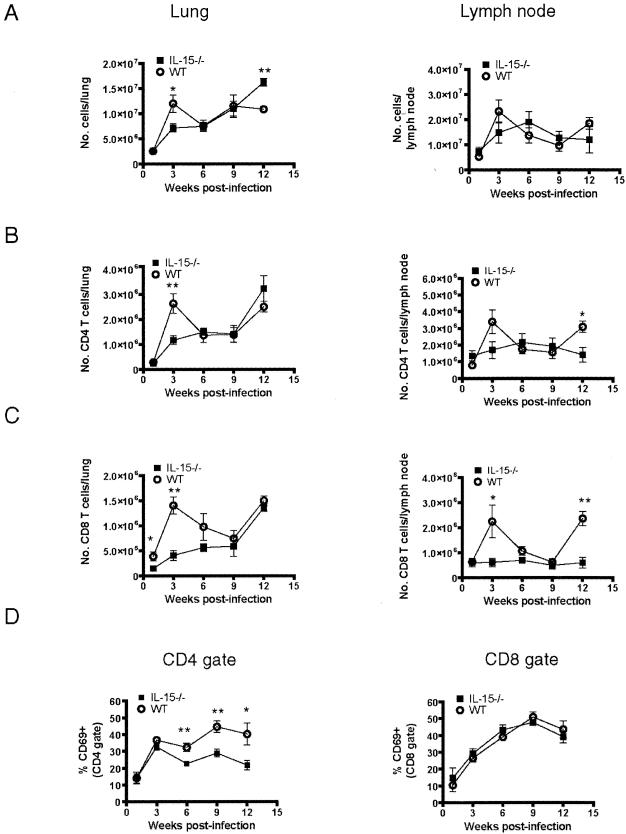
Several studies have provided evidence that interleukin-15 (IL-15) can enhance protective immune responses against Mycobacterium tuberculosis infection. However, the effects of IL-15 deficiency on the functionality of M. tuberculosis-specific CD4 and CD8 T cells are unknown. In this study, we investigated the generation and maintenance of effector and memory T-cell responses following M. tuberculosis infection of IL-15(-/-) mice. IL-15(-/-) mice had slightly higher bacterial numbers during chronic infection, which were accompanied by an increase in gamma interferon (IFN-gamma)-producing CD4 and CD8 T cells. There was no evidence of increased apoptosis or a defect in proliferation of CD8 effector T cells following M. tuberculosis infection. The induction of cytotoxic and IFN-gamma CD8 T-cell responses was normal in the absence of IL-15 signaling. The infiltration of CD4 and CD8 T cells into the lungs of "immune" IL-15(-/-) mice was delayed in response to M. tuberculosis challenge. These findings demonstrate that efficient effector CD4 and CD8 T cells can be developed following M. tuberculosis infection in the absence of IL-15 but that recall T-cell responses may be impaired.


Figures

FIG. 1.
FIG. 1.
IL-15−/− mice control primary and secondary low-dose M. tuberculosis infection. (A) IL-15−/− and wild-type mice were infected with a low dose of M. tuberculosis via the aerosol route (primary infection). (B) Prolonged antibiotic treatment, which commenced at 4 weeks after primary infection and lasted for 5 months, ensured complete elimination of mycobacteria and establishment of a memory pool of CD4 and CD8 T cells. After a resting period, IL-15−/− and wild-type mice were challenged with a low dose of M. tuberculosis via the aerosol route (secondary infection). At designated time points, serial dilutions of lung, spleen, and lymph node homogenates were plated on 7H10 plates, and the CFU were enumerated after 21 days of incubation. The results represent the mean ± standard error of the mean (SEM) of four mice per group.
FIG. 2.
FIG. 2.
Phenotypical characterization of immune cells infiltrating the lungs of IL-15−/− and wild-type mice. (A) Overall numbers of live cells inside the lungs and lymph nodes of IL-15−/− and wild-type mice were determined by the trypan blue exclusion method. (B and C) CD4 and CD8 T cells in the lungs and lymph nodes of IL-15−/− and wild-type mice were analyzed by flow cytometry gating on lymphocyte population by forward and side scatter. The absolute numbers were obtained using the following formula: total number of cells per organ × % CD4+ cells (or % CD8+ cells) within live cell gate. (D) The activation status of CD4 and CD8 T cells was assessed by staining lung cells with anti-CD4, anti-CD8, and anti-CD69 antibodies. The results are presented as the percentage of CD69-positive cells within CD4 and CD8 gates. The data are presented as the mean ± SEM of results from four mice per group, and the experiments were repeated three times. Statistical analysis was determined by a two-tailed Student t test. *, P ≤ 0.05; **, P ≤ 0.01.
FIG. 3.
FIG. 3.
CD8 T-cell effector functions following M. tuberculosis infection. (A) To estimate the proliferative capacity of CD8 T cells inside the lungs of IL-15−/− and wild-type mice, BrdU was injected intraperitoneally 16 h prior to each experiment. Single lung suspensions were stained with anti-CD8 and anti-BrdU antibodies. The results shown are the mean percentages of proliferating (BrdU+) cells within the CD8 T-cell gate ± SEM. Statistical significance was determined by a two-tailed Student t test. *, P ≤ 0.05; **, P ≤ 0.01. (B) The amount of apoptosis occurring within the CD8 T-cell population was determined after staining lung cells with anti-CD8 antibody, annexin V, and 7-AAD. The results are the percentage of annexin V- and 7-AAD-positive cells within the CD8 T-cell gate ± SEM. No significant differences were observed in the percentage of apoptotic cells within CD8 T cells of IL-15−/− and wild-type mice. (C and D) The number of IFN-γ-producing CD4 and CD8 T cells was determined by ELISPOT assay after ex vivo stimulation of lung cells with M. tuberculosis-infected dendritic cells incubated with blocking anti-MHC-I or anti-MHC-II antibodies, respectively. The inhibitory effect of anti-MHC-I and anti-MHC-II antibodies was shown to be ≥95% at a 10-μg/ml concentration. The background number of SFU (<10 SFU) after incubation of lung cells with uninfected dendritic cells was subtracted before calculations were made. The number of IFN-γ-producing T cells was determined by multiplying the frequency of IFN-γ-producing CD4 or CD8 T cells by the total number of lung cells. The results shown are the mean numbers of IFN-γ-producing cells ± SEM of four mice per group. Experiments were performed twice. The statistical significance was determined by a two-tailed Student t test. *, P ≤ 0.05; **, P ≤ 0.01. (E) The frequency of CTLp in the lungs of IL-15−/− and wild-type mice was determined by LDA. Serial dilutions of lung cells (40,000 to 1,250 cells/well; 24 replicates/cell dilution) were stimulated with M. tuberculosis-infected dendritic cells for 7 days, followed by another round of stimulation with M. tuberculosis-infected macrophages. After 14 days of expansion, the cytotoxicity of each individual well was determined by chromium release assay using M. tuberculosis-infected macrophages as targets. Spontaneous lysis was determined in 24 replicates containing chromium-labeled macrophages in media without effector T cells. The wells were considered to be positive for cytotoxicity if the amount of chromium released was greater than the mean spontaneous lysis plus 3 standard deviations. The actual frequency of CTLp was determined by χ2 minimization analysis. The experiments were repeated twice with similar results.
FIG. 4.
FIG. 4.
Cytokine profile of IL-15−/− mice. RNA was isolated from the lungs of IL-15−/− and wild-type mice at designated time points. Lung RNA from uninfected wild-type mice was used as a calibrator, and endogenous HPRT was used as a normalizer gene. Relative gene expression was calculated as 2−ΔΔCt, where ΔCt = Ct (gene of interest) − Ct (normalizer) and ΔΔCt = ΔCt (sample) − ΔCt (calibrator). We used published sequences for the IL-2, IL-7, IL-10, and IL-12 primer and probe sets (5), which were used at 400 and 250 nM concentrations, respectively. The primer-probe efficiency for each set was >99%. The graphs summarize the staining of four mice per group, and statistical significance was determined by the Mann-Whitney test. *, P ≤ 0.05; **, P ≤ 0.01.
FIG. 5.
FIG. 5.
Exogenous administration of IL-15 did not significantly change the course of M. tuberculosis infection. Mice were injected intraperitoneally with recombinant human IL-15 (1 μg/mouse daily) for 21 days starting at day 5 postinfection until day 26 postinfection. (A) CFU were determined after plating lung homogenates on 7H10 plates following 21 days of incubation. (B and C) Lung single-cell suspensions were stained with anti-CD4 and anti-CD8 antibodies, and the percentage of CD4 and CD8 T cells within the lymphocyte gates was determined by flow cytometry.
FIG. 6.
FIG. 6.
Lung lymphocyte analysis after secondary infection with M. tuberculosis. Mice were infected with a low dose of M. tuberculosis via the aerosol route for 4 weeks (primary infection) prior to commencing antibiotic treatment for 5 months to ensure complete elimination of mycobacteria and establishment of a memory pool of CD4 and CD8 T cells. After a resting period, IL-15−/− and wild-type mice were challengedwith a low dose of M. tuberculosis via the aerosol route (secondary infection), and memory T-cell responses were analyzed. (A) Overall numbers of viable cells in the lungs of IL-15−/− and wild-type mice following M. tuberculosis challenge were determined by the trypan blue exclusion method. (B and C) The absolute numbers of CD4 and CD8 T cells were determined after staining lung cells with anti-CD4 and anti-CD8 antibodies. The numbers were calculated by multiplying the total number of lung cells by the percent CD4+ or CD8+ within live cell gate. The results presented are means ± SEM of four mice per group. (D) The percentage of activated CD4 and CD8 T cells in the lungs of IL-15−/− and wild-type mice was evaluated by staining for CD69 expression using flow cytometry. Data represent mean percentages of CD69+ T cells within CD4 and CD8 gates ± SEM. The graphs summarize the staining of cells from four mice per group, and statistical significance was determined by the Student t test. *, P ≤ 0.05; **, P ≤ 0.01.
FIG. 7.
FIG. 7.
Memory CD4 and CD8 T cells are not impaired in proliferation or cytokine production in the absence of IL-15 following M. tuberculosis challenge. (A) The percentage of proliferating (BrdU+) cells within CD4 and CD8 gates was determined as described in the text and the legend to Fig. 3. The graphs represent means ± SEM of results from four mice per group. Statistical differences were calculated using a two-sided Student t test. *, P ≤ 0.05; **, P ≤ 0.01. CD4 and CD8 T cells from “immune” IL-15−/− and wild-type mice showed similar proliferation kinetics. (B) The numbers of IFN-γ-producing CD4 and CD8 T cells in the lungs of “immune” IL-15−/− and wild-type mice were determined by ELISPOT using M. tuberculosis-infected dendritic cells as antigen-specific stimulators. Incubation of M. tuberculosis-infected dendritic cells with blocking MHC-I or MHC-II antibodies delineated the number of IFN-γ-producing CD4 or CD8 T cells, respectively. The inhibitory effect of the blocking MHC-I and MHC-II antibodies was ≥95% at a 10-μg/ml concentration. The calculations were made as described in the legend to Fig. 3. The mean numbers of IFN-γ-producing CD4 and CD8 T cells from four IL-15−/− and wild-type mice at each time point are shown. Error bars represent SEM.

Similar articles

Cited by

References

    1. Avice, M. N., C. E. Demeure, G. Delespesse, M. Rubio, M. Armant, and M. Sarfati. 1998. IL-15 promotes IL-12 production by human monocytes via T cell-dependent contact and may contribute to IL-12-mediated IFN-gamma secretion by CD4+ T cells in the absence of TCR ligation. J. Immunol. 161:3408-3415. - PubMed
    1. Becker, T. C., E. J. Wherry, D. Boone, K. Murali-Krishna, R. Antia, A. Ma, and R. Ahmed. 2002. Interleukin 15 is required for proliferative renewal of virus-specific memory CD8 T cells. J. Exp. Med. 195:1541-1548. - PMC - PubMed
    1. Berard, M., K. Brandt, S. Bulfone-Paus, and D. F. Tough. 2003. IL-15 promotes the survival of naive and memory phenotype CD8+ T cells. J. Immunol. 170:5018-5026. - PubMed
    1. Doherty, T. M., R. A. Seder, and A. Sher. 1996. Induction and regulation of IL-15 expression in murine macrophages. J. Immunol. 156:735-741. - PubMed
    1. Giulietti, A., L. Overbergh, D. Valckx, B. Decallonne, R. Bouillon, and C. Mathieu. 2001. An overview of real-time quantitative PCR: applications to quantify cytokine gene expression. Methods 25:386-401. - PubMed

Publication types

MeSH terms

LinkOut - more resources