. 2005 May;73(5):3025-37.
doi: 10.1128/IAI.73.5.3025-3037.2005.

Relapsing fever spirochetes contain chromosomal genes with unique direct tandemly repeated sequences

Affiliations

Relapsing fever spirochetes contain chromosomal genes with unique direct tandemly repeated sequences

Cyril Guyard et al. Infect Immun. 2005 May.

Abstract

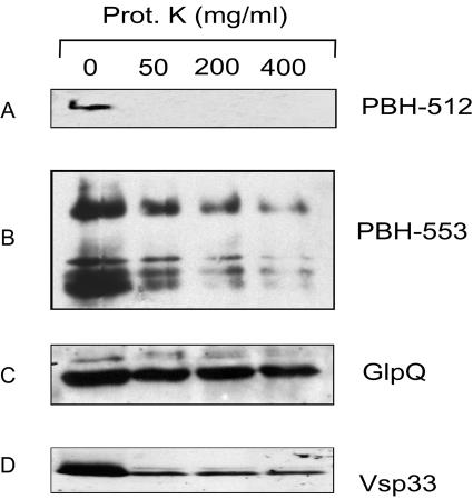
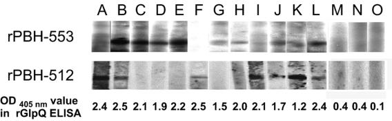
Genome sequencing of the relapsing fever spirochetes Borrelia hermsii and Borrelia turicatae identified three open reading frames (ORFs) on the chromosomes that contained internal, tandemly repeated amino acid sequences that were absent in the Lyme disease spirochete Borrelia burgdorferi. The predicted amino acid sequences of these genes (BH0209, BH0512, and BH0553) have hydrophobic N termini, indicating that these proteins may be secreted. B. hermsii transcribed the three ORFs in vitro, and the BH0512- and BH0553-encoded proteins (PBH-512 and PBH-553) were produced in vitro and in experimentally infected mice. PBH-512 and PBH-553 were on the spirochete's outer surface, and antiserum to these proteins reduced the adherence of B. hermsii to red blood cells. PCR analyses of 28 isolates of B. hermsii and 8 isolates of B. turicatae demonstrated polymorphism in each gene correlated with the number of repeats. Serum samples from relapsing fever patients reacted with recombinant PBH-512 and PBH-553, suggesting that these proteins are produced during human infection. These polymorphic proteins may be involved in the pathogenicity of these relapsing fever spirochetes and provide a mechanism for antigenic heterogeneity within their populations.


Figures

FIG. 1.
FIG. 1.
Illustration of the three chromosomal genes, BH0209 (A), BH0512 (B), and BH0553 (C), and respective hydropathy profiles of their deduced amino acid sequences using the Kyte and Doolittle algorithm (18). In the large arrows at the top, the locations of DNA coding the unique repeat domains and putative signal sequences are represented by gray and black boxes, respectively. Putative signal sequences, deduced amino acid sequences, and the respective number of repeats are shown below the large arrows. Broken lines represent regions omitted from the diagram of BH0512 (7,185 bp) to shorten its schematic length. Hydropathy profiles were determined using a window size of nine amino acids. The vertical axis displays relative hydrophobicity with negative scores indicating relative hydrophilicity. The horizontal axis indicates amino acid numbers. Thin single-headed arrows indicate hydrophobic N termini (N-ter); thin double-headed arrows indicate the repeat regions of the proteins.
FIG. 2.
FIG. 2.
Polymorphism analyses of BH0209 (A), BH0512 (B), and BH0553 (C) in 28 isolates of B. hermsii (as labeled with their codes above each gel). After PCR amplification, the products were electrophoresed in a 1% agarose gel and visualized after ethidium bromide staining. The lengths of the amplification products were estimated using Quantity One software. The positions of the molecular size markers (M) are indicated on both sides of the gels in bp.
FIG. 3.
FIG. 3.
Alignments of repeated domains encoded by BH0512 from the B. hermsii isolates DAH, SIL, COM, and CMC. Black boxes with white type represent amino acid residues conserved in the four isolates, and white boxes with boldface type represent amino acid residues present in at least 50% of the sequences.
FIG. 4.
FIG. 4.
Polymorphism analyses of BT0209 (A), BT0512 (B), and BT0553 (C) in eight isolates of B. turicatae (as labeled above each gel). After PCR amplification, the products were electrophoresed in a 1% agarose gel and visualized after ethidium bromide staining. The lengths of the amplification products were estimated using Quantity One software. The positions of the molecular size markers (M) are indicated on both sides of the gels in bp.
FIG. 5.
FIG. 5.
Immunoblot analyses of B. hermsii DAH, B. turicatae 91E135, and B. burgdorferi B31 probed with PBH-512 antibodies (A) and PBH-553 antibodies (B). Comparative immunoblot analyses of B. hermsii DAH lysates produced in vitro and in vivo using PBH-512 antibodies (C) and PBH-553 antibodies (D). Primary bound antibodies were detected with horseradish peroxidase-linked goat anti-rabbit IgG. The positions of the molecular mass markers are indicated on the left of each immunoblot in kDa.
FIG. 6.
FIG. 6.
Localization of B. hermsii PBH-512 and PBH-553. Immunoblot analyses of B. hermsii cells treated or not treated with proteinase K (Prot. K) by use of specific antibodies to detect removal of PBH-512 (A) and PBH-553 (B). Control proteins GlpQ (periplasmic) (C) and Vsp33 (surface exposed) (D) were respectively used for accessibility to proteinase K degradation. After proteinase K treatment, the amounts of PBH-512 and PBH-553 were significantly reduced. Bound antibodies were detected with horseradish peroxidase-linked goat anti-rabbit IgG.
FIG. 7.
FIG. 7.
Immunoblot analyses of 28 isolates of B. hermsii using antisera specific for PBH-512 (A) and PBH-553 (B). The codes for the isolates are indicated at the tops of the immunoblots. Brackets indicate the isolates from Genomic Groups I and II based on 16S rRNA, flaB, gyrB, and glpQ sequence analysis (26). The positions of the molecular mass markers are indicated on the left in kDa. Bound antibodies were detected with 125I-labeled protein A.
FIG. 8.
FIG. 8.
Immunoblot analyses of purified recombinant PBH-512 and PBH-553 using 12 human relapsing fever sera and 3 naïve human sera (labeled A through O above immunoblots). Seven of the human relapsing fever sera (58%) reacted with recombinant PBH-512, and eight (67%) reacted with recombinant PBH-553. All human relapsing fever sera were positive, with anti-glpQ antibodies determined by enzyme-linked immunosorbent assay (ELISA) and with rGlpQ indicated at the bottom. As negative controls, experiments were also performed with three normal human sera, none of which immunoreacted significantly with recombinant GlpQ, PBH-512, or PBH-553. Bound antibodies were detected with 125I-labeled protein A. OD, optical density.
FIG. 9.
FIG. 9.
Antibody-mediated inhibition of B. hermsii DAH adhesion to red blood cells. (A) Microphotograph of B. hermsii DAH bound to red blood cells. (B) Percentages of B. hermsii DAH bound to red blood cells after incubation with antibodies against PBH-553 or PBH-0512, control antibody (anti-flagellin), or normal serum. The percentages of bound B. hermsii cells were 46.3 and 39.1% for anti-PBH-512 serum and anti-PBH-553 serum, respectively. Error bars indicate standard deviations of the results from three separate experiments.*, Student's t test P value = 0.01 versus experiment with normal rabbit serum; **, Student's t test P value = 0.004 versus experiment with normal rabbit serum.

References

    1. Andrade, M. A., C. Perez-Iratxeta, and C. P. Ponting. 2001. Protein repeats: structures, functions, and evolution. J. Struct. Biol. 134:117-131. - PubMed
    1. Banerjee, S. N., M. Banerjee, K. Fernando, W. Burgdorfer, and T. G. Schwan. 1998. Tick-borne relapsing fever in British Columbia, Canada: first isolation of Borrelia hermsii. J. Clin. Microbiol. 36:3505-3508. - PMC - PubMed
    1. Barbour, A. G. 1984. Isolation and cultivation of Lyme disease spirochetes. Yale J. Biol. Med. 57:521-525. - PMC - PubMed
    1. Barbour, A. G., and B. I. Restrepo. 2000. Antigenic variation in vector-borne pathogens. Emerg. Infect. Dis. 6:449-457. - PMC - PubMed
    1. Breitschwerdt, E. B., W. L. Nicholson, A. R. Kiehl, C. Steers, D. J. Meuten, and J. F. Levine. 1994. Natural infections with Borrelia spirochetes in two dogs from Florida. J. Clin. Microbiol. 32:352-357. - PMC - PubMed

MeSH terms

Substances

LinkOut - more resources