

Invasion of epithelial cells by locus of enterocyte effacement-negative enterohemorrhagic Escherichia coli
- PMID: 15845514
- PMCID: PMC1087320
- DOI: 10.1128/IAI.73.5.3063-3071.2005
Invasion of epithelial cells by locus of enterocyte effacement-negative enterohemorrhagic Escherichia coli
Abstract
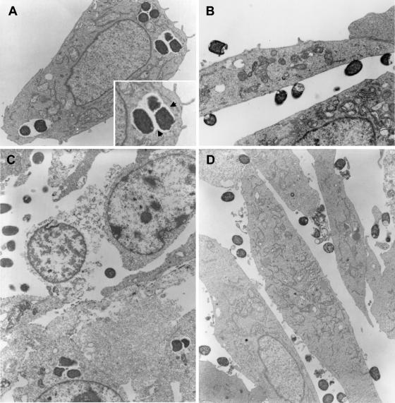
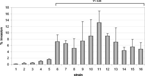
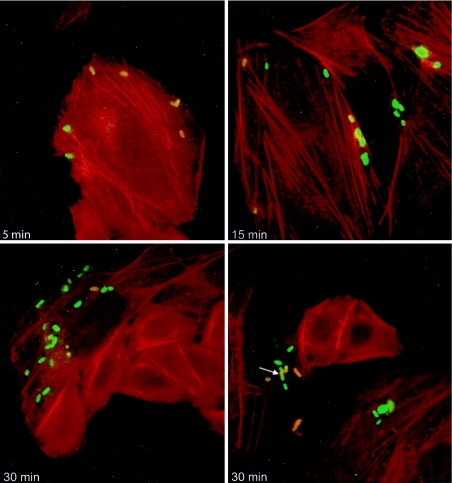
The majority of enterohemorrhagic Escherichia coli (EHEC) strains associated with severe disease carry the locus of enterocyte effacement (LEE) pathogenicity island, which encodes the ability to induce attaching and effacing lesions on the host intestinal mucosa. While LEE is essential for colonization of the host in these pathogens, strains of EHEC that do not carry LEE are regularly isolated from patients with severe disease, although little is known about the way these organisms interact with the host epithelium. In this study, we compared the adherence properties of clinical isolates of LEE-negative EHEC with those of LEE-positive EHEC O157:H7. Transmission electron microscopy revealed that LEE-negative EHEC O113:H21 was internalized by Chinese hamster ovary (CHO-K1) epithelial cells and that intracellular bacteria were located within a membrane-bound vacuole. In contrast, EHEC O157:H7 remained extracellular and intimately attached to the epithelial cell surface. Quantitative gentamicin protection assays confirmed that EHEC O113:H21 was invasive and also showed that several other serogroups of LEE-negative EHEC were internalized by CHO-K1 cells. Invasion by EHEC O113:H21 was significantly reduced in the presence of the cytoskeletal inhibitors cytochalasin D and colchicine and the pan-Rho GTPase inhibitor compactin, whereas the tyrosine kinase inhibitor genistein had no significant impact on bacterial invasion. In addition, we found that EHEC O113:H21 was invasive for the human colonic cell lines HCT-8 and Caco-2. Overall these studies suggest that isolates of LEE-negative EHEC may employ a mechanism of host cell invasion to colonize the intestinal mucosa.
Figures
Similar articles
-
Identification of a novel fimbrial gene cluster related to long polar fimbriae in locus of enterocyte effacement-negative strains of enterohemorrhagic Escherichia coli.Infect Immun. 2002 Dec;70(12):6761-9. doi: 10.1128/IAI.70.12.6761-6769.2002. Infect Immun. 2002. PMID: 12438351 Free PMC article.
-
Hyperadherence of an hha mutant of Escherichia coli O157:H7 is correlated with enhanced expression of LEE-encoded adherence genes.FEMS Microbiol Lett. 2005 Feb 1;243(1):189-96. doi: 10.1016/j.femsle.2004.12.003. FEMS Microbiol Lett. 2005. PMID: 15668018
-
Characterization of Saa, a novel autoagglutinating adhesin produced by locus of enterocyte effacement-negative Shiga-toxigenic Escherichia coli strains that are virulent for humans.Infect Immun. 2001 Nov;69(11):6999-7009. doi: 10.1128/IAI.69.11.6999-7009.2001. Infect Immun. 2001. PMID: 11598075 Free PMC article.
-
[Plasticity of bacterial genomes: pathogenicity islands and the locus of enterocyte effacement (LEE)].Berl Munch Tierarztl Wochenschr. 2004 Mar-Apr;117(3-4):116-29. Berl Munch Tierarztl Wochenschr. 2004. PMID: 15046458 Review. German.
-
Interkingdom Chemical Signaling in Enterohemorrhagic Escherichia coli O157:H7.Adv Exp Med Biol. 2016;874:201-13. doi: 10.1007/978-3-319-20215-0_9. Adv Exp Med Biol. 2016. PMID: 26589220 Review.
Cited by
-
Escherichia coli O157:H7 Curli Fimbriae Promotes Biofilm Formation, Epithelial Cell Invasion, and Persistence in Cattle.Microorganisms. 2020 Apr 17;8(4):580. doi: 10.3390/microorganisms8040580. Microorganisms. 2020. PMID: 32316415 Free PMC article.
-
The type 4 pili of enterohemorrhagic Escherichia coli O157:H7 are multipurpose structures with pathogenic attributes.J Bacteriol. 2009 Jan;191(1):411-21. doi: 10.1128/JB.01306-08. Epub 2008 Oct 24. J Bacteriol. 2009. PMID: 18952791 Free PMC article.
-
Diversity of Hybrid- and Hetero-Pathogenic Escherichia coli and Their Potential Implication in More Severe Diseases.Front Cell Infect Microbiol. 2020 Jul 15;10:339. doi: 10.3389/fcimb.2020.00339. eCollection 2020. Front Cell Infect Microbiol. 2020. PMID: 32766163 Free PMC article. Review.
-
Virulence characterization of Shiga-toxigenic Escherichia coli isolates from wholesale produce.Appl Environ Microbiol. 2011 Jan;77(1):343-5. doi: 10.1128/AEM.01872-10. Epub 2010 Nov 5. Appl Environ Microbiol. 2011. PMID: 21057025 Free PMC article.
-
Plasmids from Shiga Toxin-Producing Escherichia coli Strains with Rare Enterohemolysin Gene (ehxA) Subtypes Reveal Pathogenicity Potential and Display a Novel Evolutionary Path.Appl Environ Microbiol. 2016 Oct 14;82(21):6367-6377. doi: 10.1128/AEM.01839-16. Print 2016 Nov 1. Appl Environ Microbiol. 2016. PMID: 27542930 Free PMC article.
References
-
- Alrutz, M. A., A. Srivastava, K. W. Wong, C. D'Souza-Schorey, M. Tang, L. E. Ch'Ng, S. B. Snapper, and R. R. Isberg. 2001. Efficient uptake of Yersinia pseudotuberculosis via integrin receptors involves a Rac1-Arp 2/3 pathway that bypasses N-WASP function. Mol. Microbiol. 42:689-703. - PubMed
-
- Bennett-Wood, V. R., J. Russell, A. M. Bordun, P. D. Johnson, and R. M. Robins-Browne. 2004. Detection of enterohaemorrhagic Escherichia coli in patients attending hospital in Melbourne, Australia. Pathology 36:345-351. - PubMed
-
- Biswas, D., K. Itoh, and C. Sasakawa. 2000. Uptake pathways of clinical and healthy animal isolates of Campylobacter jejuni into INT-407 cells. FEMS Immunol. Med. Microbiol. 29:203-211. - PubMed
Publication types
MeSH terms
Substances
LinkOut - more resources
Full Text Sources
