

doi: 10.1128/IAI.73.5.3137-3146.2005.
Surface analyses and immune reactivities of major cell wall-associated proteins of group a streptococcus
Affiliations
- PMID: 15845522
- PMCID: PMC1087385
- DOI: 10.1128/IAI.73.5.3137-3146.2005
Item in Clipboard
Surface analyses and immune reactivities of major cell wall-associated proteins of group a streptococcus
Infect Immun.
2005 May.
Abstract
A proteomic analysis was undertaken to identify cell wall-associated proteins of Streptococcus pyogenes. Seventy-four distinct cell wall-associated proteins were identified, 66 of which were novel. Thirty-three proteins were immunoreactive with pooled S. pyogenes-reactive human antisera. Biotinylation of the GAS cell surface identified 23 cell wall-associated proteins that are surface exposed.
Figures

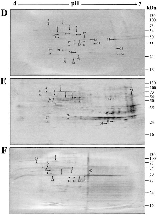
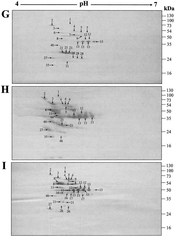
Two-dimensional gel electrophoresis profiles of GAS mutanolysin cell wall extracts. The extracts were harvested from GAS strains NS931 (A, D, and G), NS13 (B, E, and H), and S43 (C, F, and I) after growth to late stationary phase (37°C for 16 h) in Todd-Hewitt medium (Difco) supplemented with 1% (wt/vol) yeast extract without shaking. The protein extracts (170 μg) were isoelectric focused over a linear pH gradient of 4 to 7 and resolved with a 12.5% SDS-PAGE gel. (A-C) The gels were stained with colloidal Coomassie blue and destained in 1%anti-human IgG-HRP conjugate (Bio-Rad). Negative-control blots probed only with goat anti-human IgG-HRP conjugate contained no immunoreactive proteins (result not shown). (G-I) The cell surface of each strain was labeled with biotin before the mutanolysin extract was harvested. The proteins were transferred to a PVDF membrane and probed with an SA-HRP conjugate prior to development with diaminobenzidine. Negative-control blots of nonbiotinylated extracts contained no labeled proteins (result not shown). Protein spots identified by peptide mass (vol/vol) acetic acid. (D-F) The proteins were transferred to a PVDF membrane and probed with a 1:100 dilution of pooled human sera from an area of endemicity. Bound antibodies were detected using a goat fingerprinting are denoted by numbered arrows, which correspond to the proteins in Table 1. Molecular mass markers are given in kilodaltons.
Two-dimensional gel electrophoresis profiles of GAS mutanolysin cell wall extracts. The extracts were harvested from GAS strains NS931 (A, D, and G), NS13 (B, E, and H), and S43 (C, F, and I) after growth to late stationary phase (37°C for 16 h) in Todd-Hewitt medium (Difco) supplemented with 1% (wt/vol) yeast extract without shaking. The protein extracts (170 μg) were isoelectric focused over a linear pH gradient of 4 to 7 and resolved with a 12.5% SDS-PAGE gel. (A-C) The gels were stained with colloidal Coomassie blue and destained in 1%anti-human IgG-HRP conjugate (Bio-Rad). Negative-control blots probed only with goat anti-human IgG-HRP conjugate contained no immunoreactive proteins (result not shown). (G-I) The cell surface of each strain was labeled with biotin before the mutanolysin extract was harvested. The proteins were transferred to a PVDF membrane and probed with an SA-HRP conjugate prior to development with diaminobenzidine. Negative-control blots of nonbiotinylated extracts contained no labeled proteins (result not shown). Protein spots identified by peptide mass (vol/vol) acetic acid. (D-F) The proteins were transferred to a PVDF membrane and probed with a 1:100 dilution of pooled human sera from an area of endemicity. Bound antibodies were detected using a goat fingerprinting are denoted by numbered arrows, which correspond to the proteins in Table 1. Molecular mass markers are given in kilodaltons.
Two-dimensional gel electrophoresis profiles of GAS mutanolysin cell wall extracts. The extracts were harvested from GAS strains NS931 (A, D, and G), NS13 (B, E, and H), and S43 (C, F, and I) after growth to late stationary phase (37°C for 16 h) in Todd-Hewitt medium (Difco) supplemented with 1% (wt/vol) yeast extract without shaking. The protein extracts (170 μg) were isoelectric focused over a linear pH gradient of 4 to 7 and resolved with a 12.5% SDS-PAGE gel. (A-C) The gels were stained with colloidal Coomassie blue and destained in 1%anti-human IgG-HRP conjugate (Bio-Rad). Negative-control blots probed only with goat anti-human IgG-HRP conjugate contained no immunoreactive proteins (result not shown). (G-I) The cell surface of each strain was labeled with biotin before the mutanolysin extract was harvested. The proteins were transferred to a PVDF membrane and probed with an SA-HRP conjugate prior to development with diaminobenzidine. Negative-control blots of nonbiotinylated extracts contained no labeled proteins (result not shown). Protein spots identified by peptide mass (vol/vol) acetic acid. (D-F) The proteins were transferred to a PVDF membrane and probed with a 1:100 dilution of pooled human sera from an area of endemicity. Bound antibodies were detected using a goat fingerprinting are denoted by numbered arrows, which correspond to the proteins in Table 1. Molecular mass markers are given in kilodaltons.
Similar articles
-
Proteomic analysis and identification of Streptococcus pyogenes surface-associated proteins.J Bacteriol. 2007 Mar;189(5):1514-22. doi: 10.1128/JB.01132-06. Epub 2006 Dec 1. J Bacteriol. 2007. PMID: 17142387 Free PMC article.
-
Characterization of a virulence-associated and cell-wall-located DNase of Streptococcus pyogenes.Microbiology (Reading). 2010 Jan;156(Pt 1):184-190. doi: 10.1099/mic.0.031955-0. Epub 2009 Oct 22. Microbiology (Reading). 2010. PMID: 19850619
-
Isolation and solubilization of gram-positive bacterial cell wall-associated proteins.Methods Mol Biol. 2008;425:295-311. doi: 10.1007/978-1-60327-210-0_24. Methods Mol Biol. 2008. PMID: 18369905
-
[Mechanisms and manifestations of population variability in Streptococcus pyogenes].Vestn Akad Med Nauk SSSR. 1986;(7):10-7. Vestn Akad Med Nauk SSSR. 1986. PMID: 2428180 Review. Russian. No abstract available.
-
Spatial positioning of cell wall-anchored virulence factors in Gram-positive bacteria.Curr Opin Microbiol. 2012 Dec;15(6):715-23. doi: 10.1016/j.mib.2012.10.010. Epub 2012 Nov 7. Curr Opin Microbiol. 2012. PMID: 23141759 Review.
Cited by
-
Vaccinology in the genome era.J Clin Invest. 2009 Sep;119(9):2515-25. doi: 10.1172/JCI38330. J Clin Invest. 2009. PMID: 19729849 Free PMC article. Review.
-
Development of a multicomponent vaccine for Streptococcus pyogenes based on the antigenic targets of IVIG.J Infect. 2016 Apr;72(4):450-9. doi: 10.1016/j.jinf.2016.02.002. Epub 2016 Feb 12. J Infect. 2016. PMID: 26880087 Free PMC article.
-
A decade of molecular pathogenomic analysis of group A Streptococcus.J Clin Invest. 2009 Sep;119(9):2455-63. doi: 10.1172/JCI38095. J Clin Invest. 2009. PMID: 19729843 Free PMC article. Review.
-
Proteome analysis of the hyaluronic acid-producing bacterium, Streptococcus zooepidemicus.Proteome Sci. 2009 Mar 28;7:13. doi: 10.1186/1477-5956-7-13. Proteome Sci. 2009. PMID: 19327162 Free PMC article.
-
Immunization against Clostridium perfringens cells elicits protection against Clostridium tetani in mouse model: identification of cross-reactive proteins using proteomic methodologies.BMC Microbiol. 2008 Nov 11;8:194. doi: 10.1186/1471-2180-8-194. BMC Microbiol. 2008. PMID: 19000325 Free PMC article.
References
-
- Altin, J. G., and E. B. Pagler. 1995. A one-step procedure for biotinylation and chemical cross-linking of lymphocyte surface and intracellular membrane-associated molecules. Anal. Biochem. 224:382-389. - PubMed
-
- Aziz, R. K., M. J. Pabst, A. Jeng, R. Kansal, D. E. Low, V. Nizet, and M. Kotb. 2004. Invasive M1T1 group A streptococcus undergoes a phase-shift in vivo to prevent proteolytic degradation of multiple virulence factors by SpeB. Mol. Microbiol. 51:123-134. - PubMed
-
- Burnette, W. N. 1981. Western blotting: electrophoretic transfer of proteins from sodium dodecyl sulfate-polyacrylamide gels to unmodified nitrocellulose and radiographic detection with antibody and radioiodinated protein A. Anal. Biochem. 112:195-203. - PubMed
-
- Chhatwal, G. S. 2002. Anchorless adhesins and invasins of Gram-positive bacteria: a new class of virulence factors. Trends Microbiol. 10:205-208. - PubMed
Publication types
MeSH terms
Substances
LinkOut - more resources
Full Text Sources
Other Literature Sources
