. 2005 Jul 1;566(Pt 1):29-35.
doi: 10.1113/jphysiol.2005.082552. Epub 2005 Apr 21.

Navigation in space--the role of the macaque ventral intraparietal area

Affiliations

Navigation in space--the role of the macaque ventral intraparietal area

Frank Bremmer. J Physiol. .

Abstract

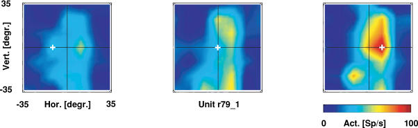
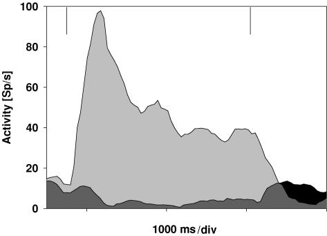
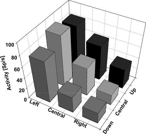
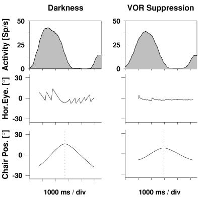
Goal-directed self-motion through space is anything but a trivial task. What we take for granted in everyday life requires the complex interplay of different sensory and motor systems. On the sensory side most importantly a target of interest has to be localized relative to one's own position in space. On the motor side the most critical step in neural processing is to define and perform a movement towards the target as well as the avoidance of obstacles. Furthermore, the multisensory (visual, tactile and auditory) motion signals as induced by one's own movement have to be identified and differentiated from the real motion of visual, tactile or auditory objects in the outside world. In a number of experimental studies performed in recent years we and others have functionally characterized a subregion within monkey posterior parietal cortex (PPC) that appears to be well suited to contribute to such multisensory encoding of spatial and motion information. In this review I will summarize the most important experimental findings on the functional properties of this very region in monkey PPC, i.e. the ventral intraparietal area.


Figures

Figure 1
Figure 1. Receptive field maps for varying eye positions
The position of each colour-coded map (red: high neural activity (spikes s−1); blue: low neural activity) represents the fixation location (given by the ‘+’ sign) during receptive field (RF) mapping. Maps are shown in screen coordinates. While the gaze shifts from left to right, the RF stays stationary on the screen, i.e. this neurone encodes visual information along the horizontal axis in head-centred coordinates. In addition, response strength increases for more rightward fixation locations.
Figure 2
Figure 2. Distribution of spatial reference frames
As described in more detail in the main text we determined for each individual neurone its reference frame for visual spatial information along the horizontal and vertical axis (see Fig. 1). Neurones whose visual RF shifted completely with the eye were considered eye-centred. Those neurones whose visual RF did not shift at all with the eye were considered head-centred. Interestingly, a number of neurones fell in an intermediate class that is neither eye- nor head-centred.
Figure 3
Figure 3. Neuronal responses for expansion and contraction stimuli
The spike density curves show the data for testing a cell with stimuli simulating forward (expansion; light grey) and backward (contraction; dark grey and black) motion. The vertical lines indicate stimulus on- and offset. The cell clearly preferred simulated forward motion.
Figure 4
Figure 4. Tuning for heading direction at the single cell level
Each column indicates the mean response for a stimulus with the singularity (i.e. heading direction) in a given part of the visual field. Heading directions were either straight ahead (central column) or 25 deg in the periphery. This cell clearly preferred heading to the left.
Figure 5
Figure 5. Responses to rotational vestibular stimulation
The upper two spike density curves show the responses of a single cell to rotational vestibular stimulation in darkness with free eye movements (left) and during VOR suppression (right). The middle panels show sample horizontal eye position traces. The bottom panels depict the position of the horizontal turntable. This cell responded to rightward motion, irrespective of whether the eye movements were suppressed or not.

Comment in

  • The senses.
    Gegenfurtner K, Kros CJ. Gegenfurtner K, et al. J Physiol. 2005 Jul 1;566(Pt 1):5. doi: 10.1113/jphysiol.2005.090837. Epub 2005 May 26. J Physiol. 2005. PMID: 15919707 Free PMC article. No abstract available.

Similar articles

Cited by

References

    1. Andersen RA, Mountcastle VB. The influence of the angle of gaze upon the excitability of the light-sensitive neurons of the posterior parietal cortex. J Neurosci. 1983;3:532–548. - PMC - PubMed
    1. Boussaoud D, Bremmer F. Gaze effects in the cerebral cortex: reference frames for space coding and action. Exp Brain Res. 1999;128:170–180. - PubMed
    1. Boussaoud D, Jouffrais C, Bremmer F. Eye position effects on the neuronal activity of dorsal premotor cortex in the macaque monkey. J Neurophysiol. 1998;80:1132–1150. - PubMed
    1. Bremmer F. Eye position effects in macaque area V4. Neuroreport. 2000;11:1277–1283. - PubMed
    1. Bremmer F, Distler C, Hoffmann KP. Eye position effects in monkey cortex. II. Pursuit- and fixation-related activity in posterior parietal areas LIP and 7A. J Neurophysiol. 1997a;77:962–977. - PubMed

Publication types

MeSH terms

LinkOut - more resources