Comparative Study
. 2005 May;25(1):6-21.
doi: 10.1002/hbm.20129.

A comparison of label-based review and ALE meta-analysis in the Stroop task

Affiliations
Comparative Study

A comparison of label-based review and ALE meta-analysis in the Stroop task

Angela R Laird et al. Hum Brain Mapp. 2005 May.

Abstract

Meta-analysis is an important tool for interpreting results of functional neuroimaging studies and is highly influential in predicting and testing new outcomes. Although traditional label-based review can be used to search for agreement across multiple studies, a new function-location meta-analysis technique called activation likelihood estimation (ALE) offers great improvements over conventional methods. In ALE, reported foci are modeled as Gaussian functions and pooled to create a statistical whole-brain image. ALE meta-analysis and the label-based review were used to investigate the Stroop task in normal subjects, a paradigm known for its effect of producing conflict and response inhibition due to subjects' tendency to perform word reading as opposed to color naming. Both methods yielded similar activation patterns that were dominated by response in the anterior cingulate and the inferior frontal gyrus. ALE showed greater involvement of the anterior cingulate as compared to that in the label-based technique; however, this was likely due to the increased spatial level of distinction allowed with the ALE method. With ALE, further analysis of the anterior cingulate revealed evidence for somatotopic mapping within the rostral and caudal cingulate zones, an issue that has been the source of some conflict in previous reviews of the anterior cingulate cortex.


Figures

Figure 1
Figure 1
Stroop foci. Activation foci of the studies included in the Stroop meta‐analyses are seen here in Talairach space in the BrainMap Search&View environment. Selected contrasts within the 19 studies yielded a total of 205 foci. A diffuse pattern of activation is seen throughout the brain in the three orthogonal views, although some convergence is evident in the cingulate cortex (sagittal view). Each color/symbol combination identifies a study within the BrainMap database, and the number displayed along with each focus refers to the experiment within the corresponding article.
Figure 2
Figure 2
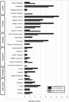
Author‐label review of the Stroop task. Author labels were recorded and tallied for all 205 foci in the Stroop articles. Labels were anatomical (“inferior frontal”), directional (“lateral frontal”), or functional (“sensorimotor”). Agreement was observed in the bilateral anterior cingulate, left inferior frontal, left inferior parietal, and left middle frontal regions.
Figure 3
Figure 3
Atlas‐label review of the Stroop task. Using a standard nomenclature reduced the number of labels from 37 (authors) to 27 (atlas). The atlas‐label review found concordance in bilateral inferior frontal gyrus, bilateral cingulate gyrus, left middle frontal gyrus, left inferior parietal lobule, and left precuneus.
Figure 4
Figure 4
Foci in the anterior cingulate or cingulate gyrus. All 205 Stroop coordinates were input to the Talairach Daemon and assigned an anatomical label. Twenty‐six foci were labeled as anterior cingulate or cingulate gyrus (listed in Table III) and are seen here along with the boundaries of the corresponding Talairach regions, viewed at slice x = 1 mm (encompassing coordinates from x = −24 to +18 mm). Most foci were located in the anterior cingulate gyrus (BA 32; green shaded region above).
Figure 5
Figure 5
Foci mislabeled by authors. Eight coordinates were labeled as cingulate gyrus or anterior cingulate gyrus by the authors, but considered mislabeled by the Talairach Daemon (listed in Table IV). The coordinates are viewed at x = 1 mm (encompassing coordinates from x = −22 to +28 mm). The shaded region in which they are plotted corresponds to the anatomical label assigned by the Talairach Daemon.
Figure 6
Figure 6
ALE maps for pooled, verbal, and manual Stroop (P < 0.01). A: In the pooled Stroop ALE meta‐analysis (19 studies, 205 foci) significant activation likelihood was seen in the anterior cingulate (z = 28, 38, 44). A lesser degree was seen in the left inferior frontal junction (z = 28, 38), the left inferior frontal gyrus (z = 10), and the left inferior parietal lobule (z = 38, 44). B: ALE meta‐analysis of verbal Stroop (13 studies, 153 foci) also revealed high ALE values in the anterior cingulate (z = 28, 38, 44) and left inferior frontal gyrus (z = 10). C: ALE meta‐analysis of manual Stroop (6 studies, 52 foci) resulted in clusters in the left inferior frontal junction (z = 28, 38, 44) and left inferior parietal lobule (z = 44).
Figure 7
Figure 7
Composite maps for verbal and manual Stroop (P < 0.05). ALE meta‐analysis of the Stroop task with a verbal response (red), manual response (blue), and the overlap between the two response types (yellow) reveals a large region of overlap within the anterior cingulate gyrus, and two smaller regions of overlap in the left inferior frontal junction (x = −46, z = 34) and left inferior parietal lobule (z = 44). Our results suggest that these three regions are major components of a network for response conflict resolution in the Stroop task.
Figure 8
Figure 8
Backsampling the ALE anterior cingulate gyrus cluster. A region of interest (ROI) was drawn around the center of mass of the ALE cluster in the anterior cingulate for the pooled Stroop meta‐analysis (left; P < 0.05) and an ROI search in BrainMap Search&View determined that 20 foci from the original studies that were located within this boundaries of this anterior cingulate gyrus ROI (right; coordinates listed in Table VI).
Figure 9
Figure 9
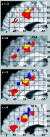
Somatotopy in the cingulate motor area. ALE meta‐analysis maps (P < 0.05) are shown for the Stroop task for verbal responses (red), manual responses (blue), and their overlap (yellow) at four slices in the sagittal orientation. A high probability for activation was found in regions that extend along the length of the cingulate sulcus, rostral to the vertical plane passing through the anterior commissure (VCA), that clearly display multiple distinct areas for verbal and manual response types.

Similar articles

Cited by

References

    1. Banich MT, Milham MP, Atchley R, Cohen NJ, Webb A, Wszalek T, Kramer AF, Liang Z‐P, Wright A, Shenker J, Magin R (2000): fMRI studies of Stroop tasks reveal unique roles of anterior and posterior brain systems in attentional selection. J Cogn Neurosci 12: 988–1000. - PubMed
    1. Bantick SJ, Wise RG, Ploghaus A, Clare S, Smith SM, Tracey I (2002): Imaging how attention modulates pain in humans using functional MRI. Brain 125: 310–319. - PubMed
    1. Barch DM, Braver TS, Akbudak E, Conturo T, Ollinger J, Snyder A (2001): Anterior cingulate cortex and response conflict: Effects of response modality and processing domain. Cereb Cortex 11: 837–848. - PubMed
    1. Becker JT, MacAndrew DK, Fiez JA (1999): A comment of the functional localization of the phonological storage subsystem of working memory. Brain Cogn 41: 27–38. - PubMed
    1. Bench CJ, Frith CD, Grasby PM, Friston KJ, Paulesu E, Frackowiak RSJ, Dolan RJ (1993): Investigations of the functional anatomy of attention using the Stroop test. Neuropsychologia 31: 907–922. - PubMed

Publication types

LinkOut - more resources