. 2005 Jun;126(1-2):13-20.
doi: 10.1016/j.jviromet.2005.01.016.

Virus-inducible reporter genes as a tool for detecting and quantifying influenza A virus replication

Affiliations

Virus-inducible reporter genes as a tool for detecting and quantifying influenza A virus replication

Andrew Lutz et al. J Virol Methods. 2005 Jun.

Abstract

The use of influenza A virus-inducible reporter gene segments in detecting influenza A virus replication was investigated. The RNA polymerase I promoter/terminator cassette was used to express RNA transcripts encoding green fluorescence protein or firefly luciferase flanked by the untranslated regions of the influenza A/WSN/33 nucleoprotein (NP) segment. Reporter gene activity was detected after reconstitution of the influenza A virus polymerase complex from cDNA or after virus infection, and was influenza A virus-specific. Reporter gene activity could be detected as early as 6 h post-infection and was virus dose-dependent. Inhibitory effects of antibodies or amantadine could be detected and quantified rapidly, providing a means of not only identifying influenza A virus-specific replication, but also of determining the antigenic subtype as well as antiviral drug susceptibility. Induction of virus-specific reporter genes provides a rapid, sensitive method for detecting virus replication, quantifying virus titers and assessing antiviral sensitivity as well as antigenic subtype.


Figures
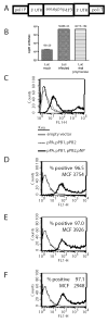
Figure 1
Figure 1
Construction and characterization of influenza A virus responsive reporter gene constructs. (A) Schematic of the FluA reporter constructs. The GFP (FluA GFP) or luciferase (FluA luc) open reading frames were cloned in place of the influenza A virus NP protein (in the negative sense, as shown by the inverted and backwards text), conserving the 5’ and 3’ untranslated regions (UTRs). The entire construct was then cloned between the human RNA polymerase I promoter (pol I P) and murine RNA polymerase I terminator (pol I T). (B) 293T cells transfected with FluA luc were mock-infected or infected with influenza A/WSN/33 (MOI=2.0). 293T cells were co-transfected with plasmids encoding the influenza polymerase proteins (PA, PB1, PB2), the nucleoprotein (NP) and the FluA luc plasmid to reconstitute the viral polymerase (Luc viral polymerase lane). 24 hours later, the cells were harvested and analyzed for luciferase activity. (C) 293T cells were transfected with FluA GFP and the indicated plasmids. 24 hours post-transfection, the cells were analyzed for GFP fluorescence by flow cytometry. MCF stands for the mean channel fluorescence of the cell population, FL 1-H is GFP fluorescence and counts indicate the number of cells with the indicated fluorescence intensity. (D-F) 293T cells were transfected with FluA GFP (thick line) or an empty vector (thin line) and infected 24 hours post-transfection with (D) A/WSN/33, (E) A/Udorn/72 or (F) A/Hong Kong/68 at an MOI of approximately 2.0. 18 hours post infection (hpi) the cells were analyzed for GFP fluorescence by flow cytometry.
Figure 2
Figure 2
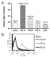
Specificity of the FluA luc and GFP constructs. (A) 293T cells were transfected with FluA luc and infected at 24 hours post-transfection with influenza A/WSN/33 (Flu A), influenza B/Yamagata/16/88 Flu B), influenza C/Jerusalem/99 (Flu C) or La Crosse virus (LAC). The cells were analyzed for luciferase activity 24 hours post infection. (B) 293T cells transfected with FluA GFP were infected at an MOI of approximately 2.0 with influenza A/WSN/33 virus (dashed line) or influenza B/Yamagata/16/88 virus (thin, solid line) or mock-infected (thick, solid line) and analyzed for GFP fluorescence by flow cytometry at 18-hpi.
Figure 3
Figure 3
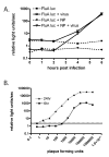
Detection limits and quantitation ranges for the FluA luc reporter gene assay. (A) 293T cells were transfected with FluA luc or FluA luc and a plasmid expressing the NP protein. 18 hours post-transfection, the cells were infected (solid lines) with 1x10⁶ PFU of influenza A/WSN/33 (MOI=2.0) or mock-infected (dashed lines) and luciferase activity assayed at the indicated times post infection. (B) Approximately 4x10⁵ 293T cells transfected with FluA luc were infected with the indicated virus inoculum and cell lysates tested for luciferase activity at 6 or 24 hours post infection. Background fluorescence levels are indicated by the dashed (24h) or solid (6h) horizontal lines.
Figure 4
Figure 4
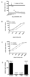
Assesing antiviral and neutralizing antibody activity using the FluA luc reporter system. (A) 293T cells transfected with FluA luc were infected with 1x10⁵ PFU of A/Udorn/72 in the presence of varying concentrations of amantadine. At 6-hpi, the cells were analyzed for luciferase activity. Luciferase activity in untreated, infected-cells is indicated by the horizontal line. (B and C) 293T cells transfected with FluA luc were infected with various PFUs of influenza A/Udorn/72 in the presence or absence of 5 μM amantadine. Cell lysates were analyzed for luciferase activity at (B) 6- or (C) 24-hpi. (D) Influenza A/WSN/33 virus (10,000 PFU, MOI=2) was incubated with the indicated dilutions of a goat anti-H1 sera for 30 minutes, then added to 293T cells transfected with the FluA luc plasmid. Six hours post infection, the cell lysates were analyzed for luciferase activity.
Figure 5
Figure 5
A stable cell line expressing a VIRGS. 293T cells were stably transfected with the FluA luc construct, infected with the indicated viruses (MOI=2.0) and luciferase activity quantified at 24 ho urs post infection.

References

    1. Azzeh M, Flick R, Hobom G. Functional analysis of the influenza A virus cRNA promoter and construction of an ambisense transcription system. Virology. 2001;289(2):400–10. - PubMed
    1. Catchpole AP, Mingay LJ, Fodor E, Brownlee GG. Alternative base pairs attenuate influenza A virus when introduced into the duplex region of the conserved viral RNA promoter of either the NS or the PA gene. J Gen Virol. 2003;84(3):507–515. - PubMed
    1. Crescenzo-Chaigne B, Naffakh N, van der Werf S. Comparative Analysis of the Ability of the Polymerase Complexes of Influenza Viruses Type A, B and C to Assemble into Functional RNPs that Allow Expression and Replication of Heterotypic Model RNA Templates In Vivo. Virology. 1999;265(2):342–353. - PubMed
    1. Ellis JS, Zambon MC. Molecular diagnosis of influenza. Rev Med Virol. 2002;12(6):375–89. - PubMed
    1. Enami M, Sharma G, Benham C, Palese P. An influenza virus containing nine different RNA segments. Virology. 1991;185(1):291–8. - PubMed

MeSH terms