. 2005 Jul;89(1):676-89.
doi: 10.1529/biophysj.104.054320. Epub 2005 Apr 22.

The dynamics and mechanics of endothelial cell spreading

Affiliations

The dynamics and mechanics of endothelial cell spreading

Cynthia A Reinhart-King et al. Biophys J. 2005 Jul.

Abstract

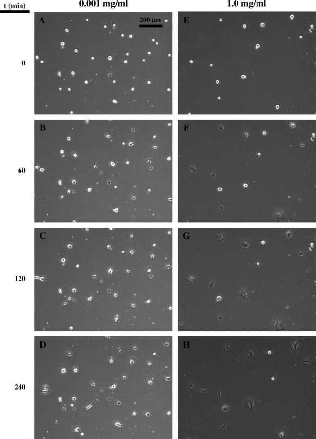
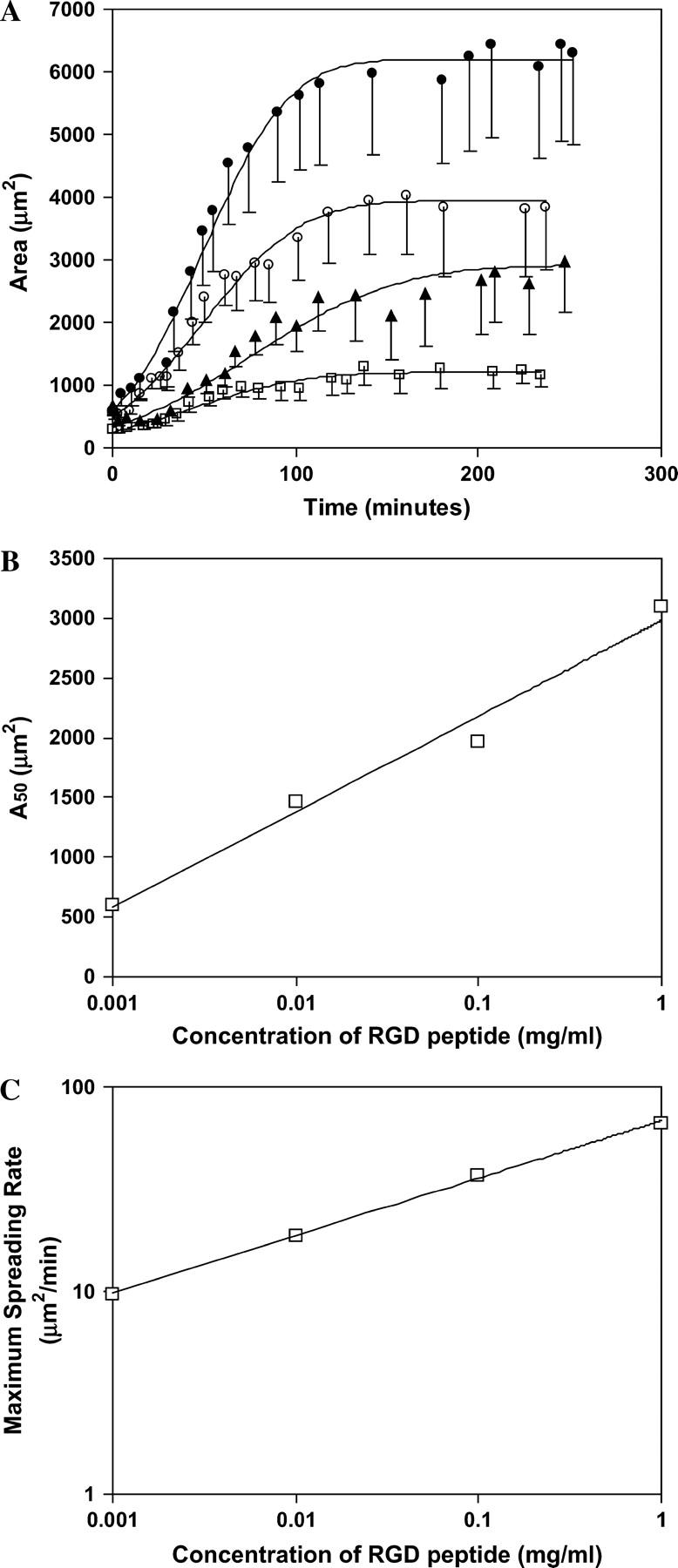
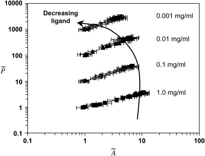
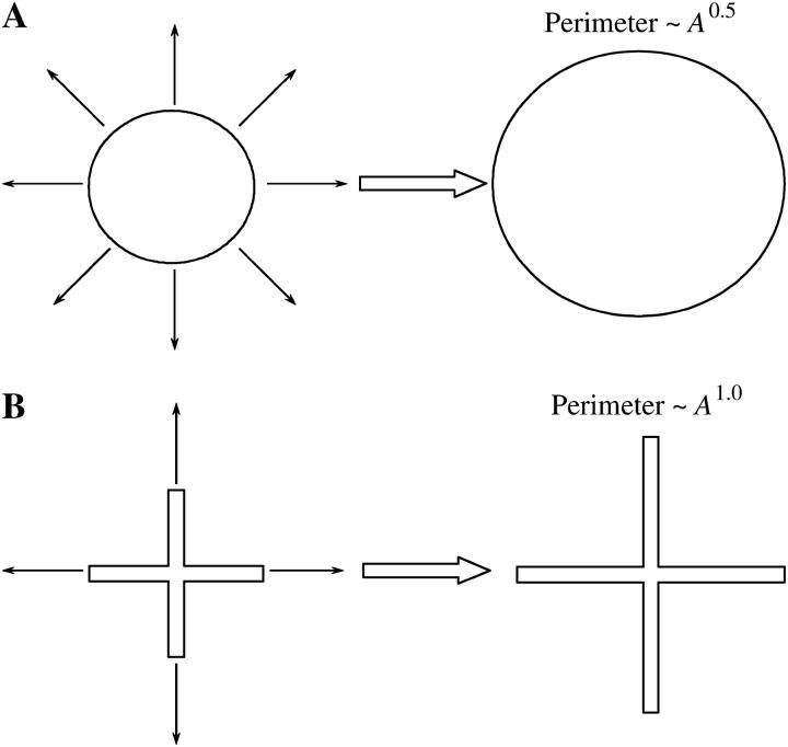
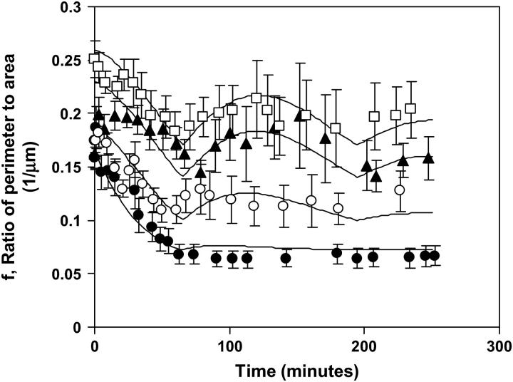
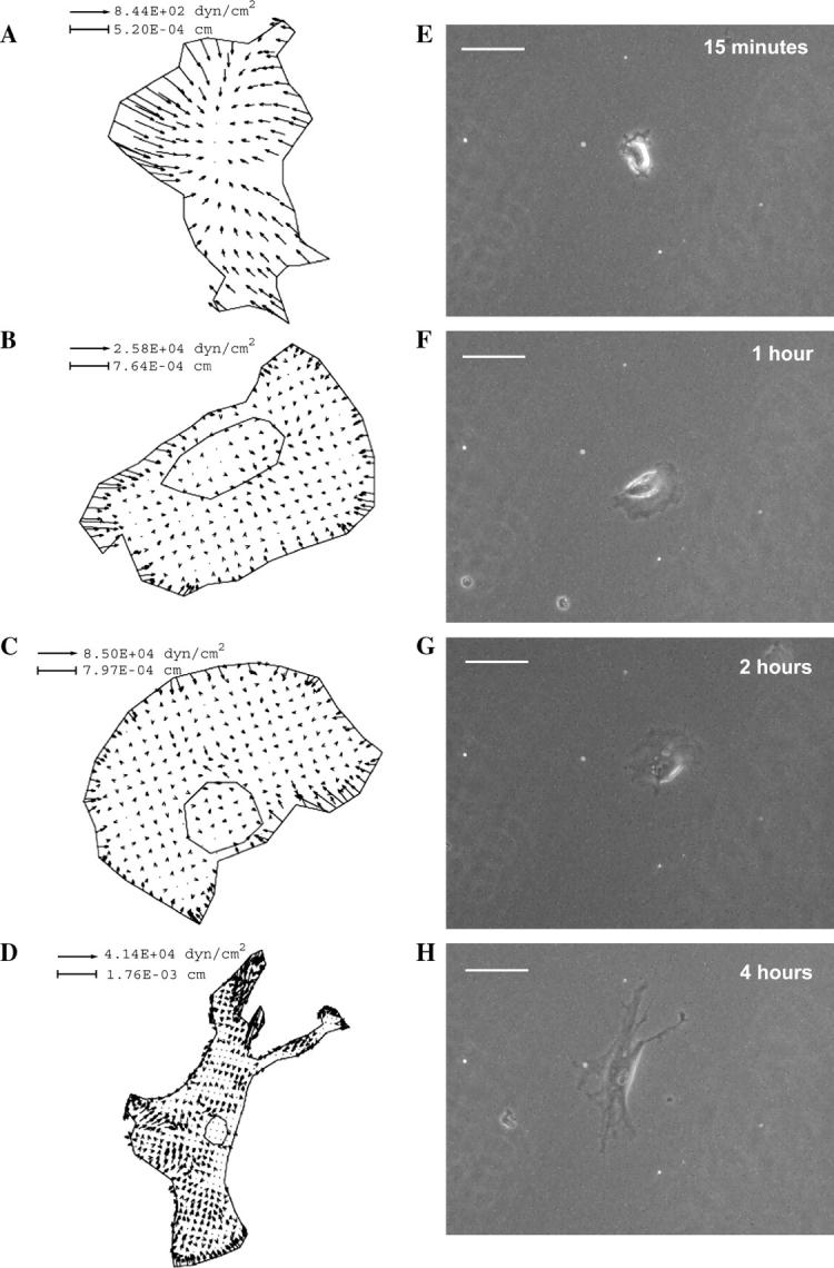
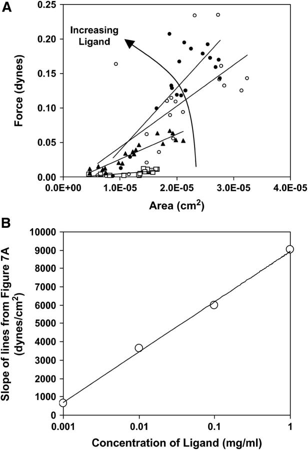
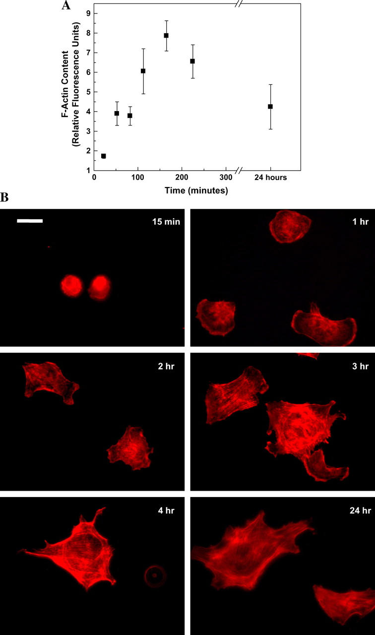
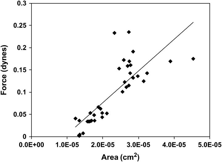
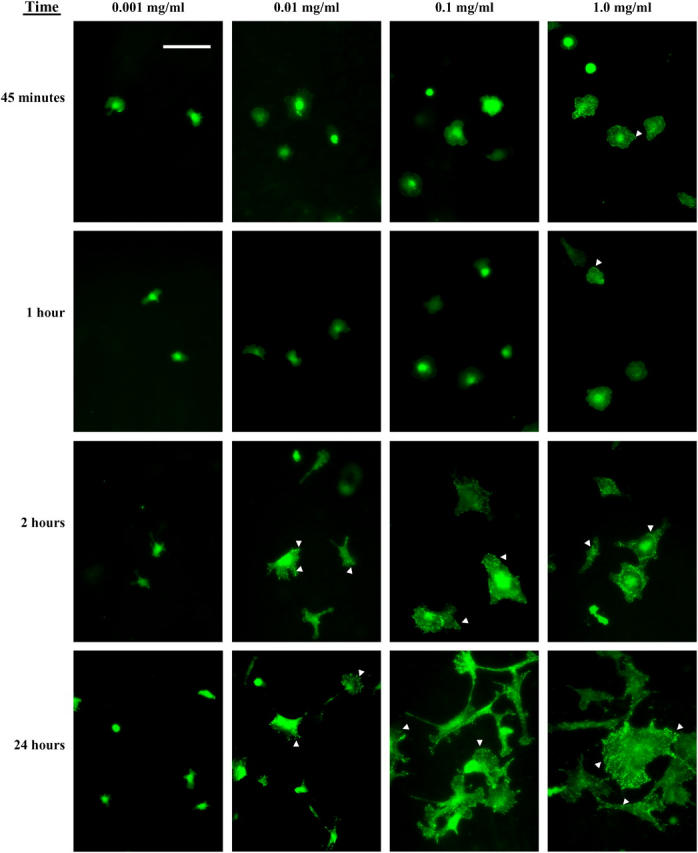
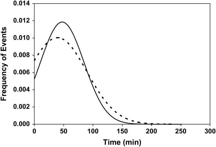
Cell adhesion to extracellular matrix is mediated by receptor-ligand interactions. When a cell first contacts a surface, it spreads, exerting traction forces against the surface and forming new bonds as its contact area expands. Here, we examined the changes in shape, actin polymerization, focal adhesion formation, and traction stress generation that accompany spreading of endothelial cells over a period of several hours. Bovine aortic endothelial cells were plated on polyacrylamide gels derivatized with a peptide containing the integrin binding sequence RGD, and changes in shape and traction force generation were measured. Notably, both the rate and extent of spreading increase with the density of substrate ligand. There are two prominent modes of spreading: at higher surface ligand densities cells tend to spread isotropically, whereas at lower densities of ligand the cells tend to spread anisotropically, by extending pseudopodia randomly distributed along the cell membrane. The extension of pseudopodia is followed by periods of growth in the cell body to interconnect these extensions. These cycles occur at very regular intervals and, furthermore, the extent of pseudopodial extension can be diminished by increasing the ligand density. Measurement of the traction forces exerted by the cell reveals that a cell is capable of exerting significant forces before either notable focal adhesion or stress fiber formation. Moreover, the total magnitude of force exerted by the cell is linearly related to the area of the cell during spreading. This study is the first to monitor the dynamic changes in the cell shape, spreading rate, and forces exerted during the early stages (first several hours) of endothelial cell adhesion.


Figures

FIGURE 1
FIGURE 1
The effect of ligand density on BAEC morphology during spreading. BAECs were plated on polyacrylamide gels containing varying amounts of RGD-containing peptide and observed under 10× magnification during spreading. Representative images from the highest and lowest densities of peptide are shown here. Cell area was significantly reduced on 0.001 mg/ml of peptide (AD), in comparison to cells on 1.0 mg/ml of peptide (EH). A and E are taken at time equal to 0; B and F are taken at time equal to 1 h; C and G are taken at time equal to 2 h; and D and H are taken at time equal to 4 h.
FIGURE 2
FIGURE 2
Ligand density controls the maximum area and spreading growth rate of spreading BAECs. (A) BAECs were plated on polyacrylamide gels containing varying amounts of RGD-containing peptide and the area of the cell was quantified and plotted against time. The results are fit to a modified error function as described in the text. (•) 1.0 mg/ml, (○) 0.1 mg/ml, (▴) 0.01 mg/ml, and (□) 0.001 mg/ml. Error bars represent mean ± SE of nine cells. (B) The area at half-maximum, as determined from the model regression in A, was plotted against the concentration of ligand on the gel, indicating a linear relationship between the log of the ligand concentration and the area of the cell. (C) The maximum growth rate, as determined from the model regression in A, was plotted against the concentration of ligand on the gel, revealing a log-linear relationship.
FIGURE 3
FIGURE 3
Ligand density controls the mode of spreading. BAECs were plated on polyacrylamide gels containing varying amounts of RGD-containing peptide and the area and perimeter of the spreading cells was quantified. The perimeter is related to the area through a power law as detailed in the text and the exponent of the power law changes with ligand density. (•) 1.0 mg/ml, (○) 0.1 mg/ml, (▴) 0.01 mg/ml, and (□) 0.001 mg/ml. Each successive ligand concentration, in decreasing order, is shifted up one decade from the next highest ligand concentration for visual clarity. Error bars represent the mean ± SE of nine cells.
FIGURE 4
FIGURE 4
(A) A schematic diagram of isotropic spreading, where the exponent in the power law equals 0.5. (B) A schematic diagram of spreading through radial projections, where the exponent approaches 1.
FIGURE 5
FIGURE 5
The ratio of perimeter/area over time exhibits oscillations at low ligand density. The ratio of perimeter/area was plotted against time for cells spreading on each of the four different ligand concentrations. Error bars represent the mean ± SE of nine cells. The data are fit to a two-parameter damped oscillator model as described in the text. As the amount of ligand increases, the number of oscillations decrease, indicating that ligand may function to support isotropic spreading and prevent pseudopodial extension. (•) 1.0 mg/ml, (○) 0.1 mg/ml, (▴) 0.01 mg/ml, and (□) 0.001 mg/ml.
FIGURE 6
FIGURE 6
Traction forces are evident early in spreading, and typically point inward throughout the entire process of spreading. Forces exerted by a cell during spreading onto its substrate were measured using traction force microscopy. (AD) Traction maps of a representative cell spreading on an intermediate concentration of peptide, 0.1 mg/ml. (EH) Phase-contrast images of those cells pictured in AE. The scale bar in the phase-contrast images is 50 μm.
FIGURE 7
FIGURE 7
Force and area are linearly related during spreading. BAECs were plated on polyacrylamide gels and immediately monitored for traction forces during spreading. Both force and area are monitored over a 4-h time period beginning at the time of plating. (A) During the first 4 h of spreading, the force exerted by the cell onto its substrate increases linearly with increasing area. (•) 1.0 mg/ml, (○) 0.1 mg/ml, (▴) 0.01 mg/ml, and (□) 0.001 mg/ml. (B) The slope of the regression lines from A were plotted against the log of the concentration of ligand, revealing a linear relationship between the two. R2 of the best fit equals 0.998.
FIGURE 8
FIGURE 8
Actin polymerization increases during spreading with a maximum at ∼3 h. (A) Cells were plated on polyacrylamide gels derivatized with 1.0 mg/ml of RGD-containing peptide and fixed at various time points. The cells were stained with Rhodamine-phalloidin, and f-actin content was quantified by comparing relative fluorescence intensities of the cells. Data are plotted with their mean ± SE. Actin polymerization increases with cell spreading and reaches a maximum at ∼3 h. By 24 h, the amount of f-actin has decreased to a steady-state value. (B) The cells were visualized using fluorescence microscopy at 60× magnification.
FIGURE 9
FIGURE 9
The force exerted by cells 3 h after plating on various densities of peptide was plotted against the cell area. The relationship between force and area approaches a steady-state value after complete actin polymerization. Three hours was chosen because it was the time at which actin polymerization peaks and stress fibers are detected. The plot reveals an approximately linear relationship with a slope of 7100 dyn/cm2. This slope is statistically similar to that found within our previous study of the same cells 24 h after plating (17), indicating that stress fibers may dominate the steady-state relationship between force and area found in our previous work.
FIGURE 10
FIGURE 10
Focal adhesions, indicated by arrowheads, form sooner on higher densities of peptide. BAECs were plated on polyacrylamide gels derivatized with varying concentration of RGD-containing peptide. At various times after plating, the cells were fixed and stained with an anti-vinculin antibody and a secondary fluorescein-conjugated antibody. The cells were then visualized at 20× magnification. Focal adhesions form much more rapidly and are much more numerous at higher concentrations of peptide. On the lowest concentration of peptide, very few focal adhesions are visible even at 24 h. The scale bar is 100 μm.
FIGURE 11
FIGURE 11
Changes in area over time during spreading are mediated by a Gaussian distribution of cell-substrate binding events. Because the changes in cell area over time during spreading can be fit to the error function, the derivative of the regression analysis to Fig. 2 was taken, illustrating a Gaussian distribution of events over time during spreading. The distribution for the highest (1.0 mg/ml, solid line) and the lowest (0.001 mg/ml, dashed line) are presented. We hypothesize that these events are the net number of binding events between receptor and substrate ligand. In this scenario, binding is most likely to occur when the cell is sufficiently spread to contact a large area of ligand, forming new bonds, but before an increase in the number of breakage events.

References

    1. Zhu, X., and R. K. Assoian. 1995. Integrin-dependent activation of MAP kinase: a link to shape-dependent cell proliferation. Mol. Biol. Cell. 6:273–282. - PMC - PubMed
    1. Ingber, D. E. 1990. Fibronectin controls capillary endothelial cell growth by modulating cell shape. Proc. Natl. Acad. Sci. USA. 87:3579–3583. - PMC - PubMed
    1. Asthagiri, A. R., C. A. Reinhart, A. F. Horwitz, and D. A. Lauffenburger. 2000. The role of transient ERK2 signals in fibronectin- and insulin-mediated DNA synthesis. J. Cell Sci. 113:4499–4510. - PubMed
    1. Gospodarowicz, D., and G. M. Lui. 1981. Effect of substrata and fibroblast growth factor on the proliferation in vitro of bovine aortic endothelial cells. J. Cell. Physiol. 109:69–81. - PubMed
    1. McAuslan, B. R., V. Bender, W. Reilly, and B. A. Moss. 1985. New functions of epidermal growth factor: stimulation of capillary endothelial cell migration and matrix dependent proliferation. Cell Biol. Int. Rep. 9:175–182. - PubMed

Publication types

LinkOut - more resources