. 2005 Apr 22;33(7):2332-42.
doi: 10.1093/nar/gki530. Print 2005.

High frequency trans-splicing in a cell line producing spliced and polyadenylated RNA polymerase I transcripts from an rDNA-myc chimeric gene

Affiliations

High frequency trans-splicing in a cell line producing spliced and polyadenylated RNA polymerase I transcripts from an rDNA-myc chimeric gene

Célia Chen et al. Nucleic Acids Res. .

Abstract

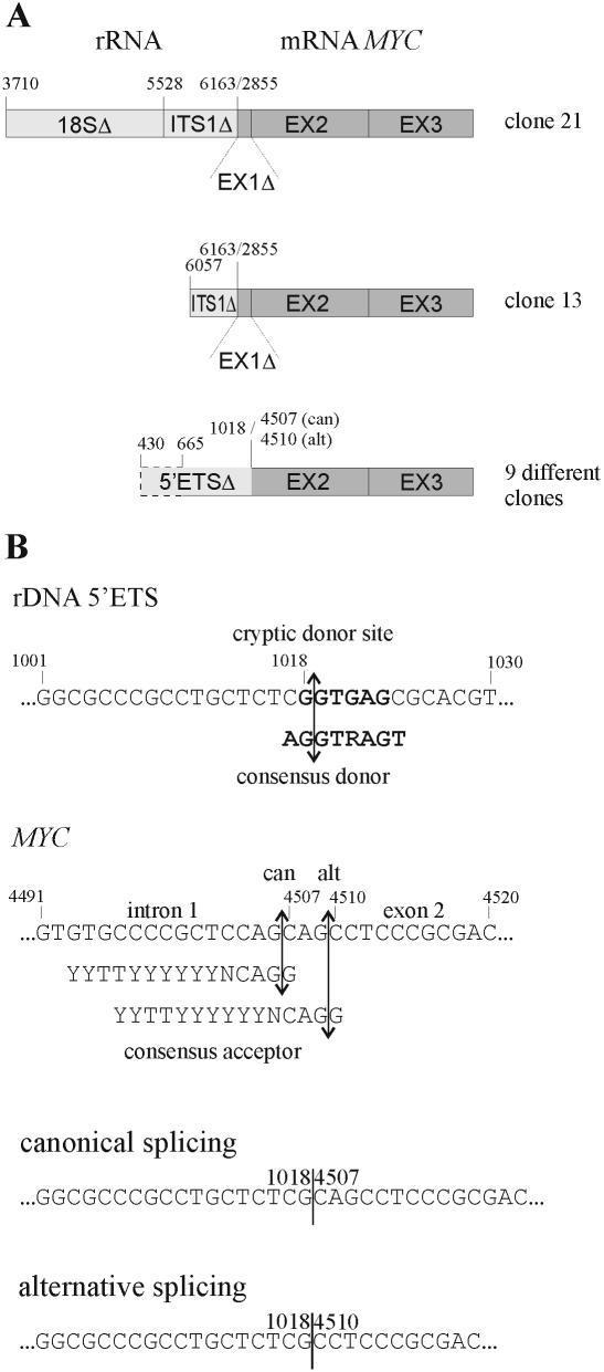
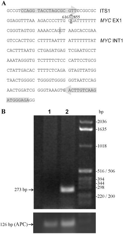
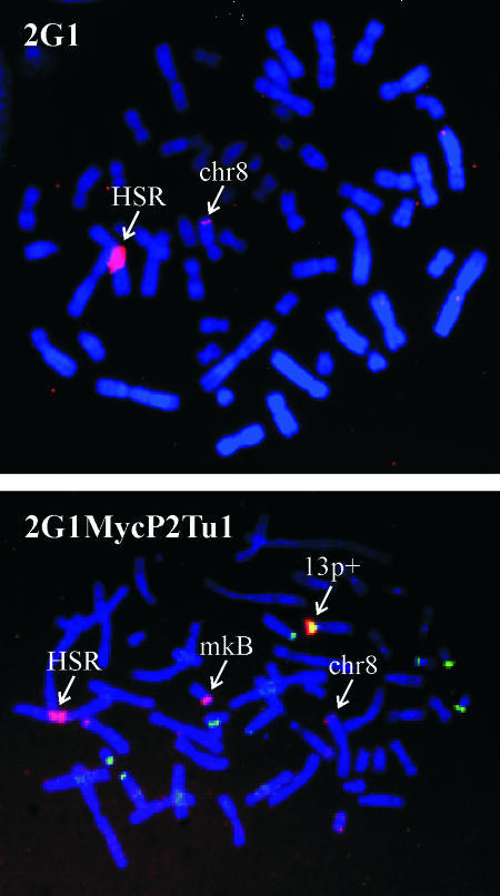
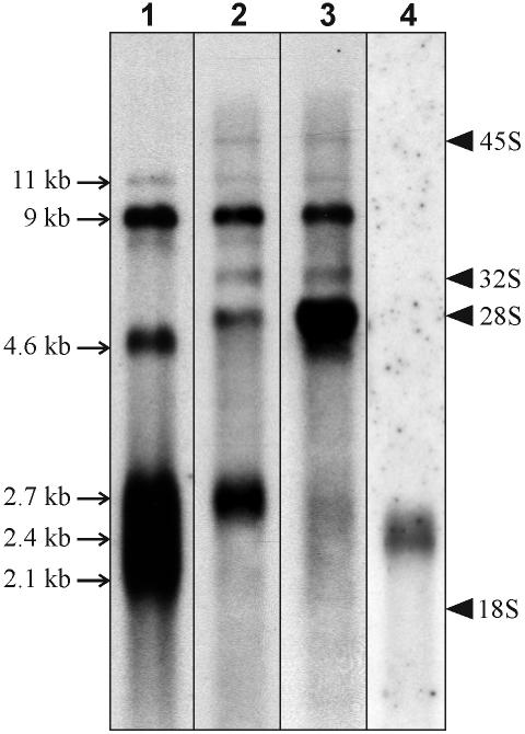
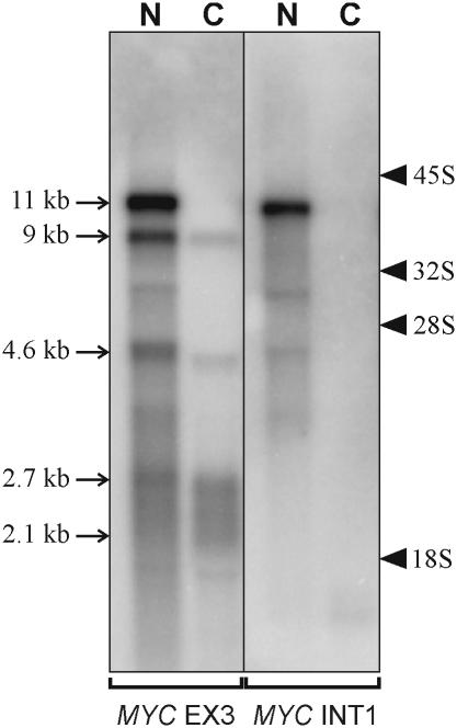
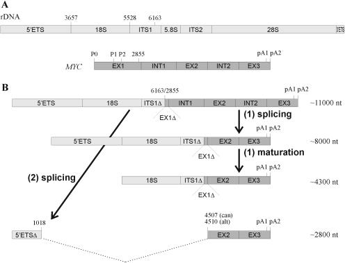
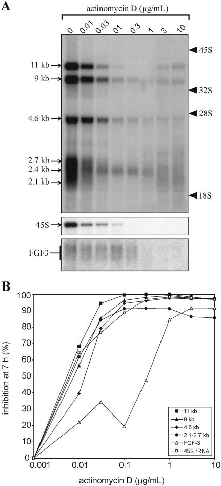
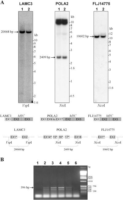
The 2G1MycP2Tu1 cell line was obtained following transfection of human colon carcinoma cells from the SW613-S cell line with a plasmid carrying a genomic copy of the human MYC gene. 2G1MycP2Tu1 cells produce MYC mRNAs and proteins of abnormal size. In order to analyze the structure of these abnormal products, a cDNA library constructed using RNA isolated from these cells was screened with a MYC probe. Fifty clones were studied by DNA sequencing. The results indicated that a truncated copy of the MYC gene had integrated into an rDNA transcription unit in 2G1MycP2Tu1 cells. This was confirmed by northern blot analysis, PCR amplification on genomic DNA and fluorescent in situ hybridization (FISH) experiments on metaphase chromosomes. 2G1MycP2Tu1 cells produce hybrid rRNA-MYC RNA molecules that are polyadenylated and processed by splicing reactions involving natural and cryptic splice sites. These transcripts are synthesized by RNA polymerase I, as confirmed by actinomycin D sensitivity experiments, suggesting that 3' end processing and splicing are uncoupled from transcription in this case. 2G1MycP2Tu1 cells also produce another type of chimeric mRNAs consisting of correctly spliced exons 2 and 3 of the MYC gene fused to one or more extraneous 5' exons by proper splicing to the acceptor sites of MYC exon 2. These foreign exons belong to 33 different genes, which are located on 14 different chromosomes. These observations and the results of FISH and Southern blotting experiments lead us to conclude that trans-splicing events occur at high frequency in 2G1MycP2Tu1 cells.


Figures

Figure 1
Figure 1
Structure of chimeric rDNA/MYC cDNAs. (A) Schematic representation of the structure of 11 cDNA clones deduced from sequencing analysis. Boxes in light and dark gray represent rDNA and MYC sequences, respectively. ITS1, internal transcribed spacer 1; 5′ETS, 5′ external transcribed sequence; EX, exon; can and alt, canonical and alternative splice acceptor site of MYC exon 2. ‘Δ’ indicates that the corresponding sequence is truncated. MYC exon1Δ is only 27 nt long. Base pair coordinates are given relative to the transcription unit for rDNA sequences (accession no. U13369) and to the genomic sequence (accession no. D10493) for MYC. The dotted line limiting the 5′ETS box between positions 430 and 665 delineates the beginning of the longest and the shortest of the nine cDNA clones. (B) Sequences of the rDNA 5′ETS and of the MYC gene encompassing the cryptic splice donor site and the acceptor site of exon 2, respectively. Consensus sequences for donor and acceptor sites are shown below, with double-headed arrows indicating the cleavage sites. The lower part of the figure shows the sequences of hybrid mRNAs after either canonical or alternative splicing.
Figure 2
Figure 2
Detection of the MYC–rDNA junction by genomic PCR. (A) DNA sequence encompassing the junction point between the MYC gene and the rDNA transcription unit. Gray arrows indicate the position of the primers used for genomic PCR. Symbols and coordinates are as in Figure 1. The junction point between ITS1 and MYC exon 1 is indicated by an I-shaped line and the border between MYC exon 1 and intron 1 by a vertical line. (B) Genomic PCR was performed on DNA extracted from 2G1 (lane 1) or 2G1MycP2Tu1 cells (lane 2). Arrows point to the 273 bp amplified fragment obtained using the primers shown in (A) and to the 126 bp fragment amplified from the APC gene. The size of the DNA fragments used as molecular weight markers is indicated on the right.
Figure 3
Figure 3
Chromosomal localization of MYC gene copies in 2G1 and 2G1MycP2Tu1 cells. FISH experiments were performed with a MYC probe (red) on 2G1 (parental cells) and 2G1MycP2Tu1 (transfectant) metaphases and with an rDNA probe (green) on 2G1MycP2Tu1 metaphases. chr8: resident MYC gene; HSR: amplified copies of the MYC gene localized in an HSR; 13p+: co-localization of the exogenous copies of the MYC gene and of rRNA genes on a rearranged chromosome 13 short arm; mkB: insertion of exogenous copies of the MYC gene into an unidentified marker chromosome. Note that the rDNA probe reveals, as expected, additional rDNA copies on other acrocentric chromosomes (green spots).
Figure 4
Figure 4
Abnormal MYC mRNAs in 2G1MycP2Tu1 cells. Polyadenylated RNA from 2G1MycP2Tu1 (lanes 1–3) or 2G1 (lane 4) cells was analyzed by northern blotting using a MYC exon 3 probe (lanes 1 and 4), a 5′ETS probe (lane 2) or an ITS1 probe (lane 3). Exposure time for lane 4 was 9 times longer than for the other lanes. Arrows on the left point to MYC mRNAs and arrowheads on the right indicate the position of rRNAs. Polyadenylated RNA preparations were not completely pure so that 45S, 32S and 28S rRNAs were visible in these samples with the rDNA probes. Detection of the 32S rRNA by the 5′ETS probe and of the 28S rRNA by the 5′ETS and ITS1 probes is due to cross-hybridization.
Figure 5
Figure 5
Nucleocytoplasmic distribution of chimeric MYC mRNAs, Northern blot analysis was performed on 10 μg of nuclear (lanes N) or cytoplasmic (lanes C) RNA extracted from 2G1MycP2Tu1 cells. An MYC exon 3 or intron 1 probe was used, as indicated. Symbols are as in Figure 4.
Figure 6
Figure 6
Possible maturation pathways for the hybrid rRNA/MYC transcripts. (A) The structures of the rDNA transcription unit and of the MYC gene are represented respectively by light and dark gray boxes. The position of the MYC gene promoters (P0, P1 and P2) and polyadenylation sites (pA1 and pA2) is indicated. (B) Two possible maturation pathways (1 and 2) are indicated by bold arrows. The approximate calculated size of the different transcripts is given on the right. 3′ETS, 3′ external transcribed spacer. Other symbols and coordinates are as in Figure 1.
Figure 7
Figure 7
Sensitivity of MYC mRNA synthesis to actinomycin D in 2G1MycP2Tu1 cells. (A) 2G1MycP2Tu1 cells were incubated for 7 h in the presence of the indicated concentrations of actinomycin D. Total cellular RNA was extracted and analyzed by northern blotting using an MYC exon 3 probe (upper panel), an rDNA 5′ETS probe which reveals the 45S ribosomal RNA (middle panel) or an FGF3 probe (lower panel). Symbols are as in Figure 4. (B) Quantification of the results shown in (A). Hybridization signals were quantified as described (30), and the results are expressed as the percentage of inhibition of the synthesis of each RNA species relative to the zero time point.
Figure 8
Figure 8
Analysis of four genomic loci in 2G1 and 2G1MycP2Tu1 cells. (A) Genomic DNA from 2G1 (lane 1) or 2G1MycP2Tu1 (lane 2) cells was analyzed by Southern blotting using a laminin gamma 3 (LAMC3) probe, a DNA polymerase alpha subunit B (POLA2) probe or a probe corresponding to the gene encoding the putative protein FLJ14775. The restriction enzyme used in each case is indicated at the bottom of each panel. Arrows point to the hybridizing fragment of interest whose size is indicated in base pairs. Note that the POLA2 probe reveals additional genomic DNA fragments since it contains several exons. The position of the DNA fragments used as molecular weight markers is indicated on the right. The structure of the corresponding chimeric cDNAs is shown with MYC exons 2 and 3 symbolized by dark gray boxes and exons from the exon-donor genes (LAMC3, POLA2 or FLJ14775) by light gray boxes. The genomic structure of these three genes in the vicinity of the exons found in the chimeric cDNAs is schematized at the bottom of the figure with the position of the restriction enzyme sites of interest and the size of the resulting fragments indicated below. The exons marked with a star are those included in the cDNA fragment used as a probe. (B) PolyA+ RNA prepared from 2G1MycP2Tu1 (lane 2), 2G1MycP2Tu1-clone 2, -clone3, -clone 4 (lanes 3–5), and 2G1 (lane 6) cells was reverse-transcribed in the presence of random hexamers. Amplification by PCR was carried out in the absence (lane 1) or in the presence (lanes 2–6) of the corresponding cDNAs. Arrow points to the 396 bp amplified fragment obtained using primers located in exon 2 of the human MSF and MYC genes. The size of the DNA fragments used as molecular weight markers is indicated on the right.

Similar articles

Cited by

References

    1. Proudfoot N.J., Furger A., Dye M.J. Integrating mRNA processing with transcription. Cell. 2002;108:501–512. - PubMed
    1. Maniatis T., Reed R. An extensive network of coupling among gene expression machines. Nature. 2002;416:499–506. - PubMed
    1. Grummt I., Skinner J.A. Efficient transcription of a protein-coding gene from the RNA polymerase I promoter in transfected cells. Proc. Natl Acad. Sci. USA. 1985;82:722–726. - PMC - PubMed
    1. Lo H.J., Huang H.K., Donahue T.F. RNA polymerase I-promoted HIS4 expression yields uncapped, polyadenylated mRNA that is unstable and inefficiently translated in Saccharomyces cerevisiae. Mol. Cell. Biol. 1998;18:665–675. - PMC - PubMed
    1. Smale S.T., Tjian R. Transcription of herpes simplex virus tk sequences under the control of wild-type and mutant human RNA polymerase I promoters. Mol. Cell. Biol. 1985;5:352–362. - PMC - PubMed

Publication types