. 2005 Jul 15;172(2):200-5.
doi: 10.1164/rccm.200502-262OC. Epub 2005 Apr 22.

Diaphragm dysfunction in chronic obstructive pulmonary disease

Affiliations

Diaphragm dysfunction in chronic obstructive pulmonary disease

Coen A C Ottenheijm et al. Am J Respir Crit Care Med. .

Abstract

Rationale: Hypercapnic respiratory failure because of inspiratory muscle weakness is the most important cause of death in chronic obstructive pulmonary disease (COPD). However, the pathophysiology of failure of the diaphragm to generate force in COPD is in part unclear.

Objectives: The present study investigated contractile function and myosin heavy chain content of diaphragm muscle single fibers from patients with COPD.

Methods: Skinned muscle fibers were isolated from muscle biopsies from the diaphragm of eight patients with mild to moderate COPD and five patients without COPD (mean FEV(1) % predicted, 70 and 100%, respectively). Contractile function of single fibers was assessed, and afterwards, myosin heavy chain content was determined in these fibers. In diaphragm muscle homogenates, the level of ubiquitin-protein conjugation was determined.

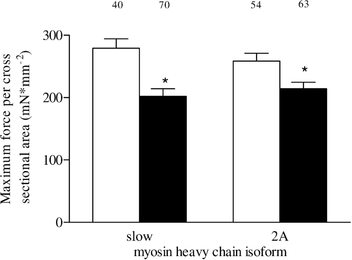
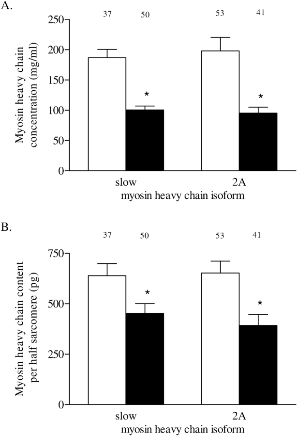
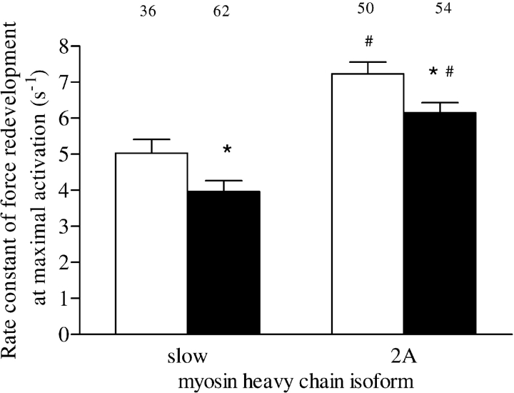
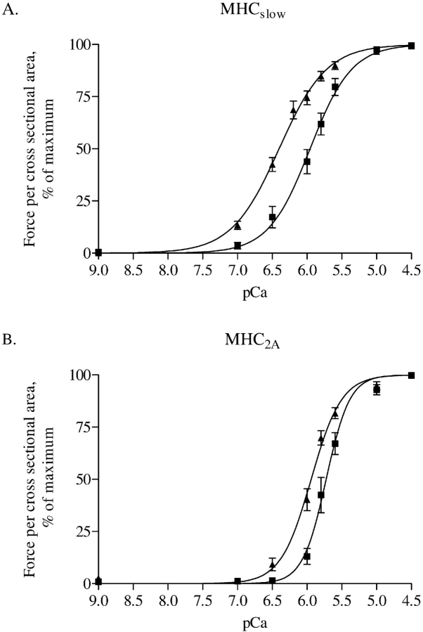
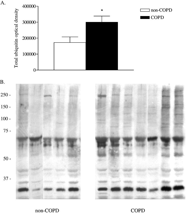
Results: Diaphragm muscle fibers from patients with COPD showed reduced force generation per cross-sectional area, and reduced myosin heavy chain content per half sarcomere. In addition, these fibers had decreased Ca2+ sensitivity of force generation, and slower cross-bridge cycling kinetics. Our observations were present in fibers expressing slow and 2A isoforms of myosin heavy chain. Ubiquitin-protein conjugation was increased in diaphragm muscle homogenates of patients with mild to moderate COPD.

Conclusions: Early in the development of COPD, diaphragm fiber contractile function is impaired. Our data suggest that enhanced diaphragm protein degradation through the ubiquitin-proteasome pathway plays a role in loss of contractile protein and, consequently, failure of the diaphragm to generate force.


Figures

<b>Figure 1.</b>
Figure 1.
Single-fiber maximum force per cross-sectional area in type slow and 2A fibers from patients with (solid bars) and without (white bars) chronic obstructive pulmonary disease (COPD). The numbers above the bars represent the total number of single fibers analyzed. Data are presented as model estimates ± SEM. *p < 0.05 difference from group without COPD.
<b>Figure 2.</b>
Figure 2.
(A) Myosin heavy chain concentration in type slow and 2A fibers from patients without COPD (white bars) and those with COPD (solid bars). Myosin heavy chain concentration was determined from sodium dodecyl sulfate–polyacrylamide gel electrophoresis densitometry of single fibers and fiber volume. (B) Myosin heavy chain content per half sarcomere in type slow and 2A fibers from patients without COPD (white bars) and those with COPD (solid bars). Myosin heavy chain content per half sarcomere was determined from single-fiber myosin heavy chain concentration and half-sarcomere volume. Myosin heavy chain content per half sarcomere estimates the number of cross bridges in parallel per single fiber. The numbers above the bars represent the total number of single fibers analyzed. Data are presented as model estimates ± SEM. *p < 0.05 difference from group without COPD.
<b>Figure 3.</b>
Figure 3.
Maximum force per half sarcomere myosin heavy chain content in type slow and 2A fibers from patients without COPD (white bars) and those with COPD (solid bars). Maximum force normalized for half-sarcomere myosin heavy chain content estimates the average force generated per cross bridge. The numbers above the bars represent the total number of single fibers analyzed. Data are presented as model estimates ± SEM.
<b>Figure 4.</b>
Figure 4.
Fraction of strongly attached cross bridges at maximal activation in type slow and 2A fibers from patients without COPD (white bars) and those with COPD (solid bars). The fraction of strongly attached cross bridges was estimated by determining the ratio of single-fiber stiffness at maximal activation in the presence of adenosine triphosphate to stiffness at maximal activation in the absence of adenosine triphosphate. The numbers above the bars represent the total number of single fibers analyzed. Data are presented as model estimates ± SEM.
<b>Figure 5.</b>
Figure 5.
Rate constant of force redevelopment at maximal activation in type slow and 2A fibers from patients without COPD (white bars) and those with COPD (solid bars). The rate constant of force redevelopment was used as a measure for cross-bridge cycling kinetics. The numbers above the bars represent the total number of single fibers analyzed. Data are presented as model estimates ± SEM. *p < 0.05 difference from group without COPD within fiber type; #p < 0.05 difference from type slow fibers within patient group.
<b>Figure 6.</b>
Figure 6.
Force–Ca2+ characteristics of single fibers from three patients without COPD and four with COPD. Isometric force generated in response to incubation of single fibers with incremental [Ca2+] was determined. Note the rightward shift of the force–Ca2+ relationship in both type slow (panel A) and 2A (panel B) single fibers from patients with COPD. Calcium concentration needed for 50% of maximal force generation was significantly higher (i.e., lower pCa50, see RESULTS) in both fiber types from patients with COPD compared with patients without COPD, indicating decreased Ca2+ sensitivity of force generation. Data are presented as model estimates ± SEM. n = 19 for patients with and without COPD, respectively, for type slow fibers; n = 10 for patients with and without COPD, respectively, for type 2A fibers. Triangles indicate patients without COPD; squares indicate patients with COPD. MHC = myosin heavy chain.
<b>Figure 7.</b>
Figure 7.
Ubiquitin–protein conjugation in diaphragm muscle homogenates of patients with COPD (n = 7) compared with the patients without COPD (n = 5). (A) Total optical density of ubiquitin–protein conjugates in diaphragm muscle homogenates. Data are presented as mean ± SEM. *p < 0.05 difference from group without COPD. (B) Antiubiquitin immunoblot of diaphragm muscle homogenates from patients with and without COPD. Each lane shows result for a diaphragm muscle homogenate from one patient. Molecular weight markers are shown in kilodaltons. Note the appearance of additional ubiquitin-protein conjugates with molecular masses between approximately 75 and 250 kD in diaphragm muscle homogenates from patients with COPD. Also, optical density of ubiquitin–protein conjugates with molecular masses of approximately 30 and approximately 70 kD, present in diaphragm muscle homogenates from patients without COPD, was increased in diaphragm muscle homogenates from patients with COPD.

Similar articles

Cited by

References

    1. Lopez AD, Murray CC. The global burden of disease, 1990–2020. Nat Med 1998;4:1241–1243. - PubMed
    1. Begin P, Grassino A. Inspiratory muscle dysfunction and chronic hypercapnia in chronic obstructive pulmonary disease. Am Rev Respir Dis 1991;143:905–912. - PubMed
    1. Zielinski J, MacNee W, Wedzicha J, Ambrosino N, Braghiroli A, Dolensky J, Howard P, Gorzelak K, Lahdensuo A, Strom K, et al. Causes of death in patients with COPD and chronic respiratory failure. Monaldi Arch Chest Dis 1997;52:43–47. - PubMed
    1. Gray-Donald K, Gibbons L, Shapiro SH, Macklem PT, Martin JG. Nutritional status and mortality in chronic obstructive pulmonary disease. Am J Respir Crit Care Med 1996;153:961–966. - PubMed
    1. Mercadier JJ, Schwartz K, Schiaffino S, Wisnewsky C, Ausoni S, Heimburger M, Marrash R, Pariente R, Aubier M. Myosin heavy chain gene expression changes in the diaphragm of patients with chronic lung hyperinflation. Am J Physiol 1998;274:L527–L534. - PubMed

Publication types

MeSH terms