

Endogenous anabolic hormones and hypermetabolism: effect of trauma and gender differences
- PMID: 15849511
- PMCID: PMC1357130
- DOI: 10.1097/01.sla.0000161028.43338.cd
Endogenous anabolic hormones and hypermetabolism: effect of trauma and gender differences
Abstract
Objective: Protein degradation, negative nitrogen balance and compromised structure of essential organs have been associated with resistance and decreased production of anabolic hormones. In turn, increased levels of anabolic hormones are associated with improved survival. The aims of the present study were to determine the pattern of anabolic hormones, resting energy expenditure and cytokines in severely thermally injured pediatric patients and to compare these parameters in female and male patients.
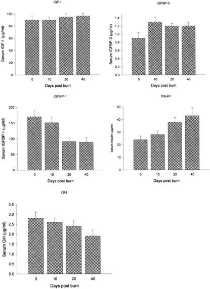
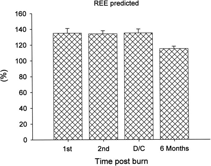
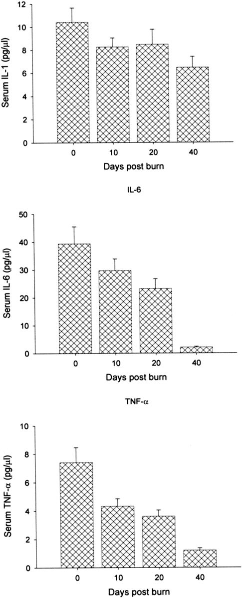
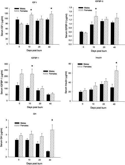
Methods: Sixty-five children (1 to 16 years of age) sustaining a severe thermal injury (> or =40% TBSA) were included into the study. Patients were further divided into females (n = 22) and males (n = 43). Patient demographics, nutritional support, incidence of sepsis, inhalation injury, and mortality were noted. Resting energy expenditure was measured during hospital course by indirect calorimetry. Blood was drawn 0, 10, 20, and 40 days postburn and serum insulin-like growth factor-I (IGF-I), insulin-like growth factor binding protein-1 and -3 (IGFBP-1, and -3), growth hormone, insulin, and cytokines were measured.
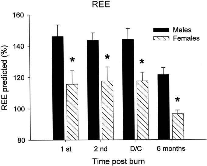
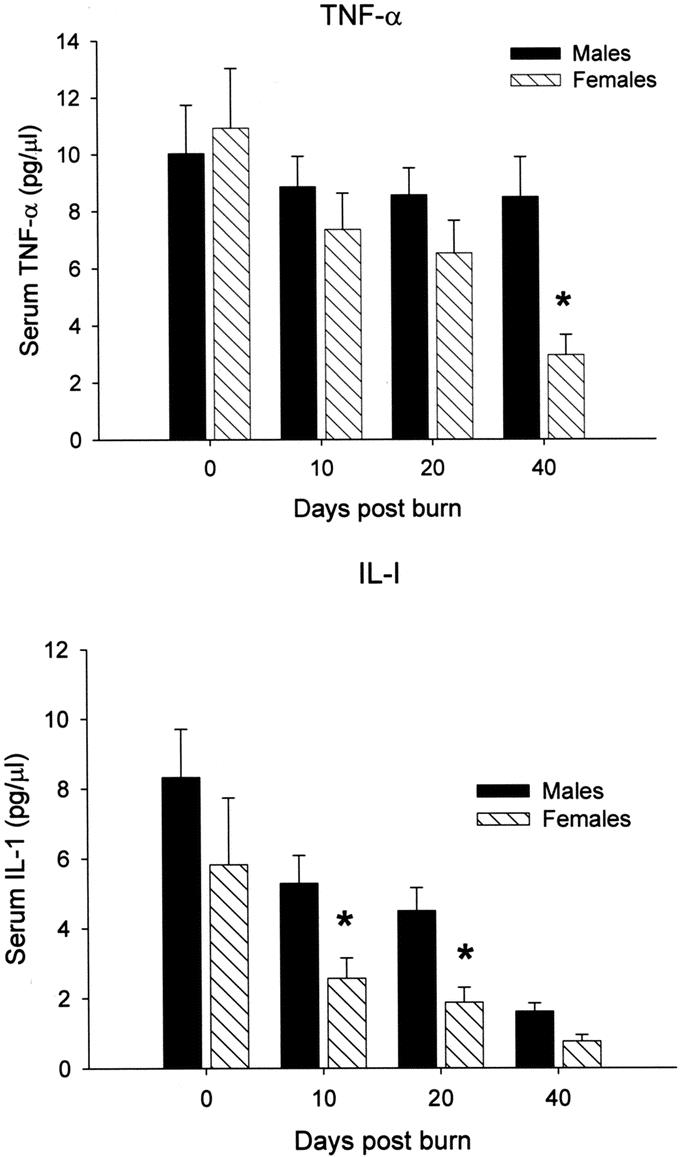
Results: There were no significant differences between females and males for demographics, nutritional intake, or concomitant injuries. In both groups, endogenous anabolic agents were drastically decreased by 3- to 5-fold up to 40 days posttrauma. Females had significantly higher levels of IGF-I, IGFBP-3, growth hormone, and insulin when compared with males, P < 0.05. Increased levels of anabolic hormones were associated with decreased stay on the ICU (females 36 +/- 22 days versus males 53+/- 39 days), decreased serum IL-1beta and TNF-alpha as well as resting energy expenditure, P < 0.05.
Conclusion: Data indicate that despite adequate nutritional support, severe thermal injury leads to decreased anabolic hormones over a prolonged period of time. Female patients had significantly increased levels of anabolic hormones, which are associated with decreased proinflammatory mediators and hypermetabolism, leading to a significant shorter ICU stay compared with male patients.
Figures
Similar articles
-
Sex differences in the long-term outcome after a severe thermal injury.Shock. 2007 May;27(5):461-5. doi: 10.1097/01.shk.0000238071.74524.9a. Shock. 2007. PMID: 17438449
-
Disturbed anabolic hormonal patterns in burned patients: the relation to glucagon.Clin Endocrinol (Oxf). 1995 Oct;43(4):491-500. doi: 10.1111/j.1365-2265.1995.tb02622.x. Clin Endocrinol (Oxf). 1995. PMID: 7586625
-
Gender differences in pediatric burn patients: does it make a difference?Ann Surg. 2008 Jul;248(1):126-36. doi: 10.1097/SLA.0b013e318176c4b3. Ann Surg. 2008. PMID: 18580216 Free PMC article.
-
Exercise training, physical fitness and the growth hormone-insulin-like growth factor-1 axis and cytokine balance.Med Sport Sci. 2010;55:128-140. doi: 10.1159/000321977. Epub 2010 Oct 14. Med Sport Sci. 2010. PMID: 20956865 Review.
-
Anabolic and anticatabolic agents used in burn care: What is known and what is yet to be learned.Burns. 2020 Feb;46(1):19-32. doi: 10.1016/j.burns.2018.03.009. Epub 2019 Dec 15. Burns. 2020. PMID: 31852612 Free PMC article. Review.
Cited by
-
Differences in Outcomes Based on Sex for Pediatric Patients Undergoing Pyloromyotomy.J Surg Res. 2020 Jan;245:207-211. doi: 10.1016/j.jss.2019.07.042. Epub 2019 Aug 14. J Surg Res. 2020. PMID: 31421364 Free PMC article.
-
Gender-specific differences in severely injured patients between 2002 and 2011: data analysis with matched-pair analysis.Crit Care. 2013 Nov 29;17(6):R277. doi: 10.1186/cc13132. Crit Care. 2013. PMID: 24289182 Free PMC article.
-
The use of anabolic agents in catabolic states.J Burns Wounds. 2007 Feb 12;6:e2. J Burns Wounds. 2007. PMID: 17364003 Free PMC article.
-
Predictors of muscle protein synthesis after severe pediatric burns.J Trauma Acute Care Surg. 2015 Apr;78(4):816-22. doi: 10.1097/TA.0000000000000594. J Trauma Acute Care Surg. 2015. PMID: 25807408 Free PMC article. Clinical Trial.
-
The use of ghrelin and ghrelin receptor agonists as a treatment for animal models of disease: efficacy and mechanism.Curr Pharm Des. 2012;18(31):4779-99. doi: 10.2174/138161212803216951. Curr Pharm Des. 2012. PMID: 22632859 Free PMC article. Review.
References
-
- Barret JP, Herndon DN. Modulation of inflammatory and catabolic responses in severely burned children by early burn wound excision in the first 24 hours. Arch Surg. 2003;138:127–132. - PubMed
-
- Takkala J, Ruokonen E, Webster NR, et al. Increased mortality associated with growth hormone treatment in critically ill adults. N Engl J Med. 1999;341:785–792. - PubMed
-
- Nguyen NT, Goldman CD, Ho HS, et al. Systemic stress response after laparoscopic and open gastric bypass. J Am Coll Surg. 2002;194:557–566. - PubMed
-
- Rennie MJ. Muscle protein turnover and wasting due to injury and disease. Br Med Bull. 1985;41:257–264. - PubMed
-
- Arnold J, Campbell IT, Samuels TA, et al. Increased whole body protein breakdown predominates over increased whole body protein synthesis in multiple organ failure. Clin Sci (Colch). 1993;84:655–661. - PubMed
Publication types
MeSH terms
Substances
LinkOut - more resources
Full Text Sources
Medical
Research Materials
Miscellaneous
