. 2005 May;115(5):1241-9.
doi: 10.1172/JCI23213. Epub 2005 Apr 21.

Targeted and restricted complement activation on acrosome-reacted spermatozoa

Affiliations

Targeted and restricted complement activation on acrosome-reacted spermatozoa

Rebecca C Riley-Vargas et al. J Clin Invest. 2005 May.

Abstract

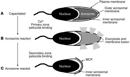
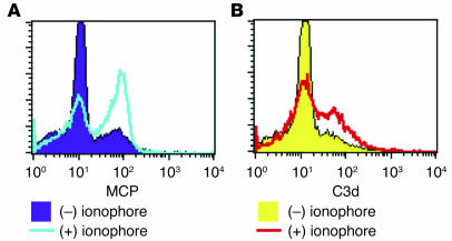
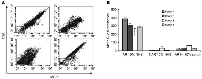
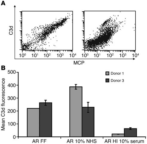
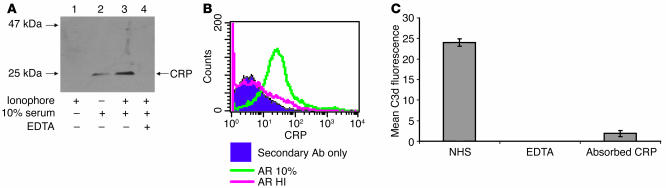
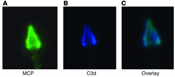
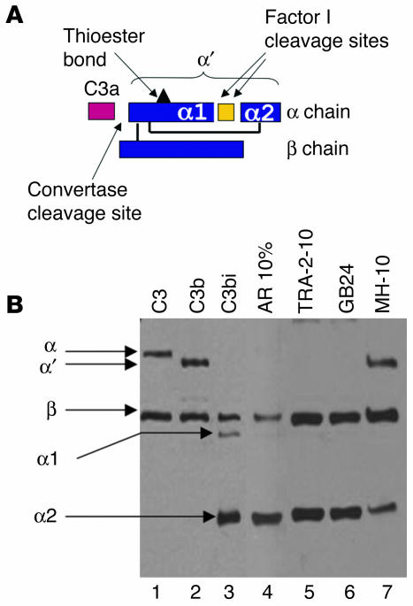
A specific hypoglycosylated isoform of the complement regulator membrane cofactor protein (MCP; CD46) is expressed on the inner acrosomal membrane (IAM) of spermatozoa. This membrane is exposed after the acrosome reaction, an exocytosis event that occurs upon contact with the zona pellucida. We initiated this investigation to assess MCP's regulatory function in situ on spermatozoa. Upon exposure of human spermatozoa to autologous serum or follicular fluid, we unexpectedly observed that acrosome-reacted spermatozoa activated the complement cascade efficiently through C3 but not beyond. Using FACS to simultaneously evaluate viability, acrosomal status, and complement deposition, we found that complement activation was initiated by C-reactive protein (CRP) and was C1q, C2, and factor B dependent. This pattern is consistent with engagement of the classical pathway followed by amplification through the alternative pathway. C3b deposition was targeted to the IAM, where it was cleaved to C3bi. Factor H, and not MCP, was the cofactor responsible for C3b cleavage. We propose that this localized deposition of complement fragments aids in the fusion process between the spermatozoa and egg, in a role akin to that of complement in immune adherence. In addition, we speculate that this "targeted and restricted" form of complement activation on host cells is a common strategy to handle modified self.


Figures

Figure 1
Figure 1
Schematic representation of the acrosome reaction of human spermatozoa. (A) The acrosome reaction of capacitated spermatozoa occurs after binding to the zona pellucida. (B) Upon this event, the outer acrosomal membrane fuses with the plasma membrane, releasing the acrosome contents. (C) MCP is expressed on the IAM and thus is exposed following the acrosome reaction.
Figure 2
Figure 2
FACS profiles of MCP and C3d on ionophore-treated spermatozoa. (A) Calcium ionophore induces the acrosome reaction, exposing MCP to the fluorescently labeled Ab. Approximately 20% of spermatozoa spontaneously undergo the acrosome reaction in the untreated sample. The antibody used was Alexa Fluor 488–labeled TRA-2-10 (mAb against MCP). (B) Spermatozoa exposed to calcium ionophore activate complement monitored by C3d deposition. The antibody used was Alexa Fluor 647–labeled mAb against C3d. A representative experiment (out of about 50 experiments) is shown.
Figure 3
Figure 3
FACS analysis of C3d deposition on viable spermatozoa exposed to 10% autologous serum. (A) Each FACS plot represents a single donor and is gated on viable spermatozoa. The correlation between the acrosome reaction (as measured by MCP staining) and complement deposition (as measured by C3d staining) is observed in all 4 donors. The inset box in each FACS plot represents the MCP- and C3d-negative population. (B) Mean C3d fluorescence of viable AR and non-AR (NAR) spermatozoa exposed to 10% normal human serum (NHS) or heat-inactivated (HI) human serum. Mean ± SD for a minimum of 3 experiments per donor is shown.
Figure 4
Figure 4
FACS analysis of viable spermatozoa exposed to follicular fluid. (A) Each FACS plot represents a single donor and is gated on viable spermatozoa. The correlation between the acrosome reaction (as measured by MCP staining) and complement deposition (as measured by C3d deposition) is observed for both donors. Each FACS plot is representative of 10 experiments. (B) Mean C3d fluorescence of viable spermatozoa exposed to follicular fluid (FF). Because of the limited quantity of neat follicular fluid, only 1 experiment using donor 1 was performed. Data for donor 3 are the mean ± SD of 3 experiments.
Figure 5
Figure 5
Analysis of CRP binding to AR spermatozoa. (A) Western blot of human spermatozoa. Ionophore-treated or untreated spermatozoa were exposed to either 10% normal or EDTA-treated autologous human serum. Lysates were separated in reducing conditions by SDS-PAGE on a 10–20% Tris-glycine gel. The primary antibody used was polyclonal goat anti–human CRP at 1:1,000 dilution. CRP is a pentamer composed of five 25-kDa subunits. (B) FACS analysis of CRP on spermatozoa. Ionophore-treated spermatozoa were exposed to either 10% normal or heat-inactivated autologous human serum. The primary antibody used was polyclonal goat anti–human CRP at 1:1,000 dilution. The secondary antibody used was FITC-linked rabbit anti-goat at 1:10,000 dilution. (C) FACS analysis of spermatozoa exposed to CRP-absorbed serum. Serum was preincubated with immobilized phosphorylcholine to absorb CRP. Spermatozoa were then exposed to 10% normal serum, 10% CRP-absorbed serum, or EDTA-treated human serum. Complement activation was measured by C3d fluorescence of viable AR spermatozoa.
Figure 6
Figure 6
Confocal microscopy of AR spermatozoa exposed to 10% serum. Spermatozoa were incubated with calcium ionophore to induce the acrosome reaction and then exposed to 10% autologous serum. (A) The mAb TRA-2-10 labeled with Alexa Fluor 488 recognizes MCP on the IAM. (B) An mAb to C3d labeled with Alexa Fluor 647 binds to the IAM. (C) Overlay of A and B. C3d deposition is limited to the IAM. Magnification, ×63.
Figure 7
Figure 7
Western blot of C3 fragments on AR spermatozoa. (A) Diagram of C3 structure. C3 is composed of 2 chains, α and β. Upon activation, C3a is released, leaving the truncated α′ chain of C3b. Factor I cleaves the α′ chain in 2 locations, creating the fragments α1, α2, and C3f (pictured in yellow). The interchain and intrachain disulfide bonds are also shown. (B) Spermatozoa were incubated with calcium ionophore to induce the acrosome reaction, exposed to 10% autologous human serum, lysed, absorbed with Sepharose linked to mAb against C3d, eluted in reducing SDS buffer, and loaded on a 10–20% gel. Lanes 1–3, C3 standards; lane 4, AR spermatozoa incubated with 10% serum; lane 5, spermatozoa were preincubated with the mAb TRA-2-10 and then exposed to 10% serum; lane 6, spermatozoa were preincubated with the mAb GB24, which blocks the complement-regulatory function of MCP; lane 7, serum was preincubated with the mAb MH-10, which blocks the complement-regulatory function of factor H (46).

Similar articles

Cited by

References

    1. Seya T, Turner J, Atkinson JP. Purification and characterization of a membrane protein (gp45-70) that is a cofactor for cleavage of C3b and C4b. J. Exp. Med. 1986;163:837–855. - PMC - PubMed
    1. Ballard LL, Bora NS, Yu GH, Atkinson JP. Biochemical characterization of membrane cofactor protein of the complement system. J. Immunol. 1988;141:3923–3929. - PubMed
    1. Seya T, Atkinson JP. Functional properties of membrane cofactor protein of complement. Biochem. J. 1989;264:581–588. - PMC - PubMed
    1. Oglesby TJ, Allen CJ, Liszewski MK, White DJ, Atkinson JP. Membrane cofactor protein (CD46) protects cells from complement-mediated attack by an intrinsic mechanism. J. Exp. Med. 1992;175:1547–1551. - PMC - PubMed
    1. Dorig RE, Marcil A, Chopra A, Richardson CD. The human CD46 molecule is a receptor for measles virus (Edmonston strain) Cell. 1993;75:295–305. - PubMed

Publication types

Substances