. 2005 May;115(5):1258-66.
doi: 10.1172/JCI24356. Epub 2005 Apr 21.

Slc11a2 is required for intestinal iron absorption and erythropoiesis but dispensable in placenta and liver

Affiliations

Slc11a2 is required for intestinal iron absorption and erythropoiesis but dispensable in placenta and liver

Hiromi Gunshin et al. J Clin Invest. 2005 May.

Abstract

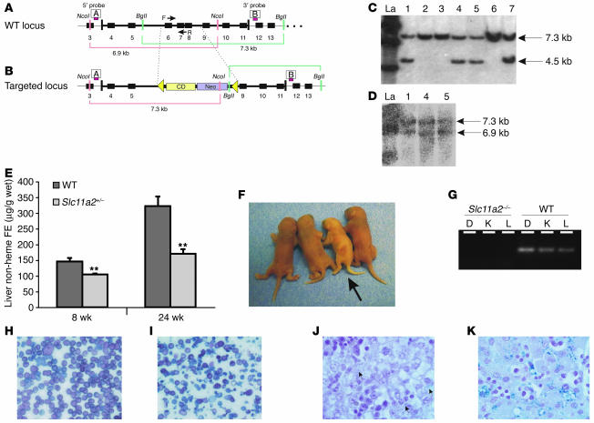
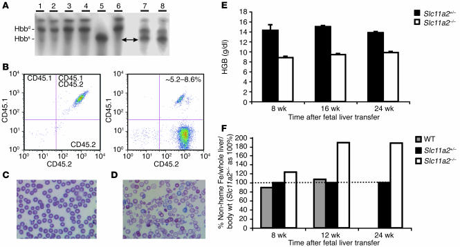
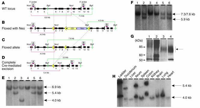
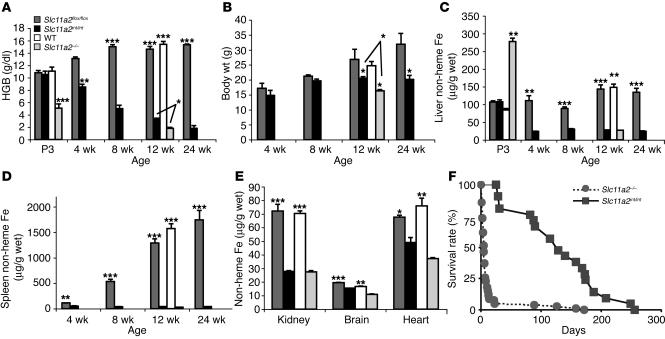
Solute carrier family 11, member 2 (SLC11A2) is the only transmembrane iron transporter known to be involved in cellular iron uptake. It is widely expressed and has been postulated to play important roles in intestinal iron absorption, erythroid iron utilization, hepatic iron accumulation, placental iron transfer, and other processes. Previous studies have suggested that other transporters might exist, but their physiological significance remained uncertain. To define the activities of Slc11a2 in vivo, we inactivated the murine gene that encodes it globally and selectively. We found that fetal Slc11a2 is not needed for materno-fetal iron transfer but that Slc11a2 activity is essential for intestinal non-heme iron absorption after birth. Slc11a2 is also required for normal hemoglobin production during the development of erythroid precursors. However, hepatocytes and most other cells must have an alternative, as-yet-unknown, iron uptake mechanism. We previously showed that Slc11a2 serves as the primary portal for intestinal iron entry in hemochromatosis. However, inactivation of murine Hfe ameliorates the phenotype of animals lacking Slc11a2.


Figures

Figure 1
Figure 1
Targeted disruption of Slc11a2. Slc11a2 wild-type locus (A) and targeted locus (B). The targeting construct contained herpes simplex virus thymidine kinase gene (HSV-TK) and neomycin-resistance (Neo) and cytidine deaminase (CD) cassettes. Homologous recombination removed exons 6–8, replacing them with neomycin-resistance and cytidine deaminase cassettes flanked by loxP sites (triangles). The 5′ and 3′ probes used for Southern blot analysis are shown as red bars labeled A and B, respectively. F, forward; R, reverse. Southern blot analysis of clones using the 3′ probe (C) and the 5′ probe (D). Clones 1, 4, and 5 were correct at both ends. La, ladder. (E) Analysis of tissue iron content in female Slc11a2+/– and wild-type mice. (F) Wild-type and Slc11a2–/– (arrow) neonates on day 1 of life. Slc11a2–/– mice were pale and runted. (G) PCR analysis of DNA prepared from duodenum (D), kidney (K), and liver (L) from Slc11a2–/– and wild-type mice using primer set F and R shown in A and B. Morphology of peripheral blood smears from wild-type (H) and Slc11a2–/– (I) mice. Original magnification, ×40. The mutant mice had hypochromic, microcytic cells with marked anisocytosis and poikilocytosis. Perls Prussian blue iron staining of wild-type neonatal liver (J) and Slc11a2–/– neonatal liver (K). Original magnification, ×100. There was little iron deposition in hepatocytes and Kupffer cells (arrows) of wild-type mice but marked iron deposition in those of Slc11a2–/– mice following a single intraperitoneal injection of iron dextran (5 mg iron).
Figure 2
Figure 2
Hematopoietic Slc11a2 is required for normal erythropoiesis. (A) Hemoglobin isoform analysis demonstrating the presence of HSC-derived hemoglobin in the peripheral blood 8 weeks after fetal liver transfer. The blood samples were as follows: lanes 1 and 2, Slc11a2–/– HSC recipients; lanes 3 and 4, wild-type HSC recipients; lane 5, C57BL/6 mice; lane 6, 129S6/SvEvTac mice; lanes 7 and 8, recipients before transfer. After fetal liver transfer, recipients produced diffuse alleles of hemoglobin β chain (Hbbd) characteristic of the 129S6/SvEvTac background but not single alleles of Hbb (Hbbs) characteristic of the C57BL/6 background. (B) Flow cytometry of peripheral blood before (left) and 8 weeks after (right) fetal liver transfer. Before HSC transfer, 100% of recipient cells had both CD45.1 and CD45.2 markers characteristic of their mixed C57BL/6 and 129S6/SvEvTac background (left). After fetal liver transfer, hematopoietic cells primarily expressed only the CD45.2 marker associated with the 129S6/SvEvTac background, which was consistent with the hemoglobin analysis. Approximately 5.2–8.6% of cells were double stained for CD45.1 and CD45.2 markers, which indicates a low level of residual chimerism. Wild-type blood morphology was normal (C) and distinctly different from that of recipients of Slc11a2–/– HSCs (D). Original magnification, ×60. The recipients of Slc11a2–/– HSCs had hypochromic, microcytic cells with anisocytosis and poikilocytosis. (E) Hemoglobin (HGB) levels were persistently lower in Slc11a2–/– HSC recipients compared with animals that received Slc11a2+/– HSCs. (F) Non-heme liver iron content (adjusted taking into account whole liver and body weight) was increased in animals that received Slc11a2–/– HSCs. Values are shown as a percent of the values determined for Slc11a2+/– HSC recipients.
Figure 3
Figure 3
Conditional inactivation of Slc11a2 in the intestine. Slc11a2 wild-type locus (A) and targeted locus (B) after introduction of an intronic floxed neomycin resistance and cytosine deaminase cassettes and an additional loxP site between exons 5 and 6. Positions of 5′ and 3′ probes used for Southern blot analysis are shown as red bars labeled A and B, respectively. (C) Partial Cre recombinase–mediated excision in which the Neo and cytosine deaminase cassettes were removed but the coding sequence was left intact. (D) Slc11a2 locus after complete excision to inactivate the gene. (E) Southern blot analysis of the 5′ end of the locus in ES cells after Cre transfection. (F) Southern blot analysis of the 3′ end of the locus in ES cells after Cre transfection. Clone 3 retained the floxed allele and was used to generate Slc11a2flox/flox mice; clones 2, 5, and 6 underwent complete deletion and were used to generate Slc11a2–/– mice without residual Neo and cytosine deaminase cassettes. (G) Immunoblot analysis of duodenum from Slc11a2–/– (lane 1), Slc11a2int/int (lane 2), wild-type (lane 3) and Trfhpx/hpx (lane 4) mice. Protein amounts loaded per gel were 4 μg of Slc11a2–/–, 2 μg of Slc11a2int/int, 50 μg of wild-type, and 2 μg of Trfhpx/hpx duodenal lysates. Slc11a2 migrates as a diffuse band between the 60-kDa and 85-kDa markers (arrow). (H) Southern blot analysis of Slc11a2int/int mice resulting from an intercross of Slc11a2flox/flox and Villin-Cre mice using probe A, demonstrating that the deletion of the floxed region occurred only in intestinal tissues.
Figure 4
Figure 4
Liver and spleen iron loading in mice with Slc11a2 mutations. (A) Hemoglobin levels. (B) Body weights. (C and D) Tissue non-heme iron content at ages 3 days (P3) to 24 weeks (E) Tissue non-heme iron content at age 12–14 weeks. Results in micrograms/gram wet weight are expressed as mean ± SEM (n = 4–16). Statistical analysis was performed using the unpaired Student’s t test, comparing mutant and wild-type mice: *P < 0.05; **P < 0.005; ***P < 0.0001. (F) Survival of mutant mice. Slc11a2–/– neonates were transfused once with rbcs on the first day of life. Slc11a2int/int mice were not transfused.

Similar articles

Cited by

References

    1. Levy JE, Jin O, Fujiwara Y, Kuo F, Andrews NC. Transferrin receptor is necessary for development of erythrocytes and the nervous system. Nat. Genet. 1999;21:396–399. - PubMed
    1. Gunshin H, et al. Cloning and characterization of a mammalian proton-coupled metal-ion transporter. Nature. 1997;388:482–488. - PubMed
    1. Canonne-Hergaux F, Gruenheid S, Ponka P, Gros P. Cellular and subcellular localization of the Nramp2 iron transporter in the intestinal brush border and regulation by dietary iron. Blood. 1999;93:4406–4417. - PubMed
    1. Canonne-Hergaux F, Zhang AS, Ponka P, Gros P. Characterization of the iron transporter DMT1 (NRAMP2/DCT1) in red blood cells of normal and anemic mk/mk mice. Blood. 2001;98:3823–3830. - PubMed
    1. Trinder D, Oates PS, Thomas C, Sadleir J, Morgan EH. Localisation of divalent metal transporter 1 (DMT1) to the microvillus membrane of rat duodenal enterocytes in iron deficiency, but to hepatocytes in iron overload. Gut. 2000;46:270–276. - PMC - PubMed

Publication types

Substances