Review
. 2004 Dec;4(4):444-65.
doi: 10.3758/cabn.4.4.444.

The primate working memory networks

Affiliations
Review

The primate working memory networks

Christos Constantinidis et al. Cogn Affect Behav Neurosci. 2004 Dec.

Abstract

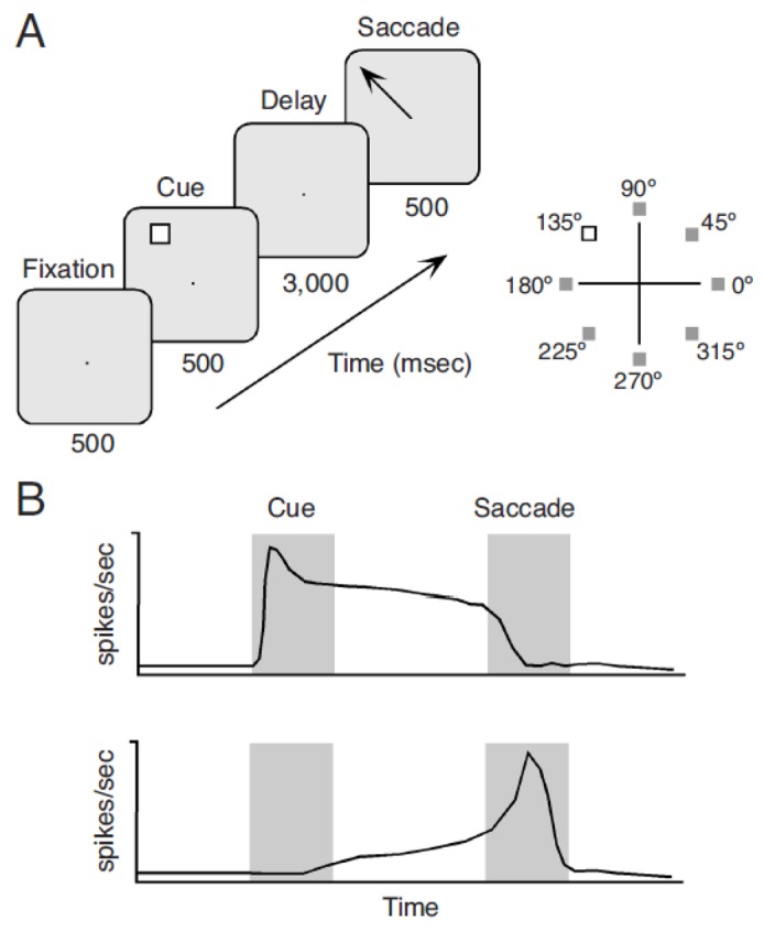
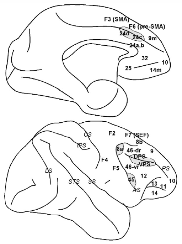
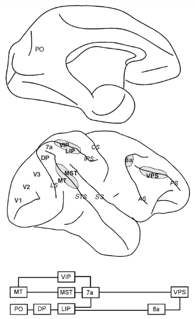
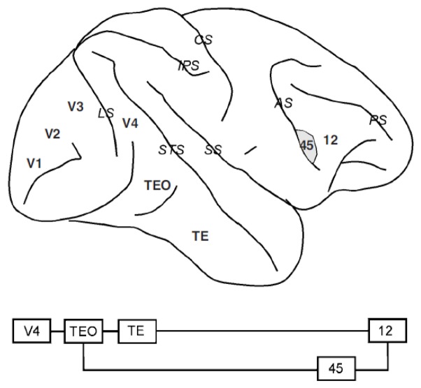
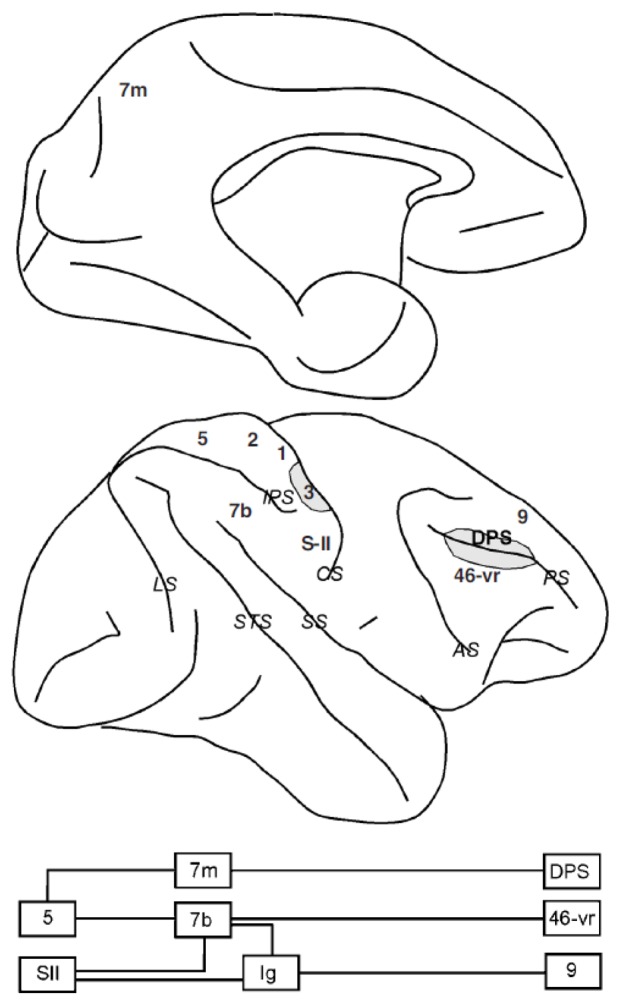
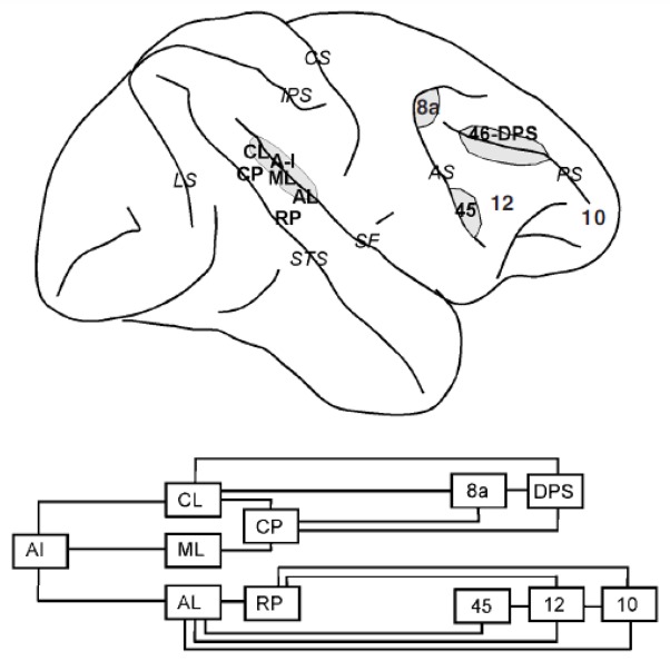
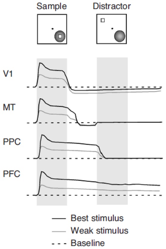
Working memory has long been associated with the prefrontal cortex, since damage to this brain area can critically impair the ability to maintain and update mnemonic information. Anatomical and physiological evidence suggests, however, that the prefrontal cortex is part of a broader network of interconnected brain areas involved in working memory. These include the parietal and temporal association areas of the cerebral cortex, cingulate and limbic areas, and subcortical structures such as the mediodorsal thalamus and the basal ganglia. Neurophysiological studies in primates confirm the involvement of areas beyond the frontal lobe and illustrate that working memory involves parallel, distributed neuronal networks. In this article, we review the current understanding of the anatomical organization of networks mediating working memory and the neural correlates of memory manifested in each of their nodes. The neural mechanisms of memory maintenance and the integrative role of the prefrontal cortex are also discussed.


Figures

Figure 1
Figure 1
(A) The ocolumotor delayed response task. Panels represent successive frames of the subject’s screen during the execution of the task. The inset on the right illustrates that the cue may appear at any of eight spatial locations around the fixation point, randomly interleaved during the experiment. (B) Schematic illustrations of two types of neuronal responses during the execution of the task. The top panel illustrates a transient response to the cue that is sustained throughout the delay period. The bottom panel shows a neuron that discharges in accelerating fashion before the onset of the eye movement. Neurons with activation at any combination of task epochs have been reported in the prefrontal cortex and elsewhere.
Figure 2
Figure 2
Schematic diagram of the macaque brain depicting prefrontal and cingulate areas. Top diagram: medial view of the brain. Bottom diagram: lateral view of the brain. Gray regions denote banks of a sulcus (only partially unfolded). Abbreviations of cortical areas: 46-dr, dorsal rim of area 46; 46-vr, ventral rim of area 46; DPS, dorsal principal sulcal area; SEF, supplementary eye field; SMA, supplementary motor area; VPS, ventral principal sulcal area. Abbreviations of sulci (in italics): AS, arcuate sulcus; CS, central sulcus; IPS, intraparietal sulcus; LS, lunate sulcus; PS, principal sulcus; SS, sylvian sulcus (lateral fissure); STS, superior temporal sulcus.
Figure 3
Figure 3
Dorsal visual pathway. Top diagram: medial view of the brain. Bottom diagram: simplified connectivity pattern between the areas of the dorsal visual pathways. Abbreviations of cortical areas: DP, dorsal parietal; LIP, lateral intraparietal; MST, medial superior temporal; MT, medial temporal; PO, parietal occipital; VIP, ventral intraparietal; VPS, ventral principal sulcal.
Figure 4
Figure 4
Ventral visual pathway. Conventions are the same as in Figure 2.
Figure 5
Figure 5
Somatosensory pathway. Abbreviations of cortical areas: Ig, insular cortex; DPS, dorsal principal sulcal; 46-vr, ventral rim of area 46.
Figure 6
Figure 6
Auditory pathways. Abbreviations of cortical areas: AL, anterior lateral; CL, caudal lateral; CP, caudal parabelt; DPS, dorsal principal sulcal; ML, middle lateral; RP, rostral parabelt.
Figure 7
Figure 7
Manifestations of memory-related activity in successive visual cortical areas. Idealized neuronal responses are shown in response to two stimuli separated by a delay period: a sample that the subject is required to remember and a subsequent distractor. The black line represents responses to an optimal stimulus evoking the best response from the neuron. The gray line represents responses to a less effective stimulus evoking a moderate neuronal response. The dotted line represents the neurons’ discharge rate baseline. Neuronal discharges in a small percentage of V1 neurons recede below the baseline during the active maintenance in memory of an optimal stimulus. Neurons in MT exhibit a short-lived persistent discharge that follows the disappearance of the stimulus. Their activity quickly returns to baseline, however. Neurons in posterior parietal cortex (PPC)—for example, in areas 7a and LIP—respond with sustained discharges in the delay period following the sample. However, these are terminated by the appearance of a distracting stimulus, outside the receptive field. Neurons in prefrontal cortex (PFC)—for example, in areas 8a and 46—respond with sustained responses to the stimulus, which are not interrupted by the appearance of the distractor.

Similar articles

Cited by

References

    1. Akkal D, Bioulac B, Audin J, Burbaud P. Comparison of neuronal activity in the rostral supplementary and cingulate motor areas during a task with cognitive and motor demands. European Journal of Neuroscience. 2002;15:887–904. - PubMed
    1. Alexander GE, DeLong MR, Strick PL. Parallel organization of functionally segregated circuits linking basal ganglia and cortex. Annual Review of Neuroscience. 1986;9:357–381. - PubMed
    1. Amador N, Schlag-Rey M, Schlag J. Reward-predicting and reward-detecting neuronal activity in the primate supplementary eye field. Journal of Neurophysiology. 2000;84(4):2166–2170. - PubMed
    1. Andersen RA, Essick GK, Siegel RM. Encoding of spatial location by posterior parietal neurons. Science. 1985;230(4724):456–458. - PubMed
    1. Andersen RA, Essick GK, Siegel RM. Neurons of area 7 activated by both visual stimuli and oculomotor behavior. Experimental Brain Research. 1987;67(2):316–322. - PubMed

Publication types