

Density of the Notch ligand Delta1 determines generation of B and T cell precursors from hematopoietic stem cells
- PMID: 15851488
- PMCID: PMC2213184
- DOI: 10.1084/jem.20042450
Density of the Notch ligand Delta1 determines generation of B and T cell precursors from hematopoietic stem cells
Abstract
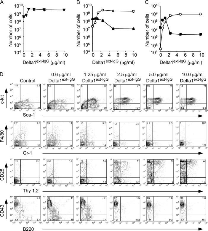
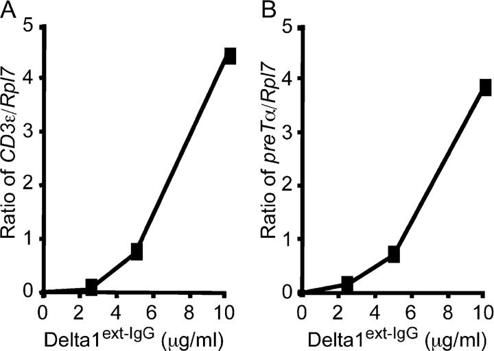
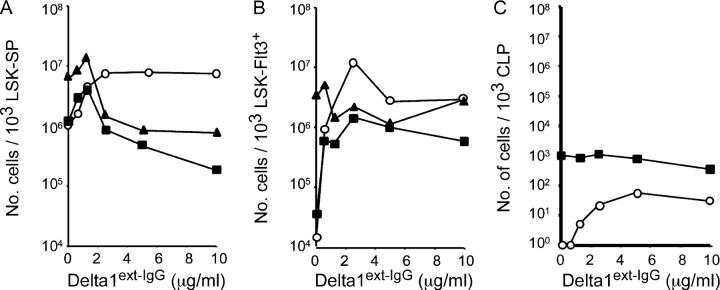
Notch signaling regulates multiple cell fate decisions by hematopoietic precursors. To address whether different amounts of Notch ligand influence lineage choices, we cultured murine bone marrow lin(-)Sca-1(+)c-kit+ cells with increasing densities of immobilized Delta1(ext-IgG) consisting of the extracellular domain of Delta1 fused to the Fc domain of human IgG1. We found that relatively lower densities of Delta1(ext-IgG) enhanced the generation of Sca-1(+)c-kit+ cells, Thy1(+)CD25+ early T cell precursors, and B220(+)CD43(-/lo) cells that, when cocultured with OP9 stroma cells, differentiated into CD19+ early B cell precursors. Higher densities of Delta1(ext-IgG) also enhanced the generation of Sca-1(+)c-kit+ precursor cells and promoted the development of Thy1(+)CD25+ cells, but inhibited the development of B220(+)CD43(-/lo) cells. Analyses of further isolated precursor populations suggested that the enhanced generation of T and B cell precursors resulted from the effects on multipotent rather than lymphoid-committed precursors. The results demonstrate the density-dependent effects of Delta1 on fate decisions of hematopoietic precursors at multiple maturational stages and substantiate the previously unrecognized ability of Delta1 to enhance the development of both early B and T precursor cells.
Figures

Similar articles
-
Enhanced T-cell reconstitution by hematopoietic progenitors expanded ex vivo using the Notch ligand Delta1.Blood. 2007 Apr 15;109(8):3579-87. doi: 10.1182/blood-2006-08-039842. Epub 2007 Jan 9. Blood. 2007. PMID: 17213287 Free PMC article.
-
Dose-dependent effects of the Notch ligand Delta1 on ex vivo differentiation and in vivo marrow repopulating ability of cord blood cells.Blood. 2005 Oct 15;106(8):2693-9. doi: 10.1182/blood-2005-03-1131. Epub 2005 Jun 23. Blood. 2005. PMID: 15976178 Free PMC article.
-
Combined effects of Notch signaling and cytokines induce a multiple log increase in precursors with lymphoid and myeloid reconstituting ability.Blood. 2003 Mar 1;101(5):1784-9. doi: 10.1182/blood-2002-06-1862. Epub 2002 Oct 31. Blood. 2003. PMID: 12411302
-
Notch signaling in lymphocyte development.Curr Opin Genet Dev. 2001 Oct;11(5):554-60. doi: 10.1016/s0959-437x(00)00232-x. Curr Opin Genet Dev. 2001. PMID: 11532398 Review.
-
Notch signalling in hematopoiesis.Semin Cell Dev Biol. 2003 Apr;14(2):143-50. doi: 10.1016/s1084-9521(02)00183-0. Semin Cell Dev Biol. 2003. PMID: 12651098 Review.
Cited by
-
Divergent effects of supraphysiologic Notch signals on leukemia stem cells and hematopoietic stem cells.Blood. 2013 Feb 7;121(6):905-17. doi: 10.1182/blood-2012-03-416503. Epub 2012 Oct 31. Blood. 2013. PMID: 23115273 Free PMC article.
-
Regulation of dendritic-cell differentiation by bone marrow stroma via different Notch ligands.Blood. 2007 Jan 15;109(2):507-15. doi: 10.1182/blood-2006-05-025601. Epub 2006 Sep 14. Blood. 2007. PMID: 16973960 Free PMC article.
-
Engineering approaches for regeneration of T lymphopoiesis.Biomater Res. 2016 Jun 29;20:20. doi: 10.1186/s40824-016-0067-1. eCollection 2016. Biomater Res. 2016. PMID: 27358746 Free PMC article. Review.
-
Notch target Hes5 ensures appropriate Notch induced T- versus B-cell choices in the thymus.Blood. 2008 Mar 1;111(5):2615-20. doi: 10.1182/blood-2007-03-079855. Epub 2007 Nov 29. Blood. 2008. PMID: 18048645 Free PMC article.
-
Epstein-Barr virus latent membrane protein 2A exploits Notch1 to alter B-cell identity in vivo.Blood. 2009 Jan 1;113(1):108-16. doi: 10.1182/blood-2008-06-160937. Epub 2008 Sep 24. Blood. 2009. PMID: 18815281 Free PMC article.
References
-
- Varnum-Finney, B., L. Xu, C. Brashem-Stein, C. Nourigat, D. Flowers, S. Bakkour, W.S. Pear, and I.D. Bernstein. 2000. Pluripotent, cytokine-dependent, hematopoietic stem cells are immortalized by constitutive Notch1 signaling. Nat. Med. 6:1278–1281. - PubMed
-
- Pui, J.C., D. Allman, L. Xu, S. DeRocco, F.G. Karnell, S. Bakkour, J.Y. Lee, T. Kadesch, R.R. Hardy, J.C. Aster, and W.S. Pear. 1999. Notch1 expression in early lymphopoiesis influences B versus T lineage determination. Immunity. 11:299–308. - PubMed
-
- Radtke, F., A. Wilson, G. Stark, M. Bauer, J. van Meerwijk, H.R. MacDonald, and M. Aguet. 1999. Deficient T cell fate specification in mice with an induced inactivation of Notch1. Immunity. 10:547–558. - PubMed
-
- Artavanis-Tsakonas, S., M.D. Rand, and R.J. Lake. 1999. Notch signaling: cell fate control and signal integration in development. Science. 284:770–776. - PubMed
Publication types
MeSH terms
Substances
Grants and funding
LinkOut - more resources
Full Text Sources
Other Literature Sources
Medical
Research Materials
Miscellaneous
