. 2005 Apr 25;169(2):219-25.
doi: 10.1083/jcb.200412019.

Disulfide bridge formation between SecY and a translocating polypeptide localizes the translocation pore to the center of SecY

Affiliations

Disulfide bridge formation between SecY and a translocating polypeptide localizes the translocation pore to the center of SecY

Kurt S Cannon et al. J Cell Biol. .

Abstract

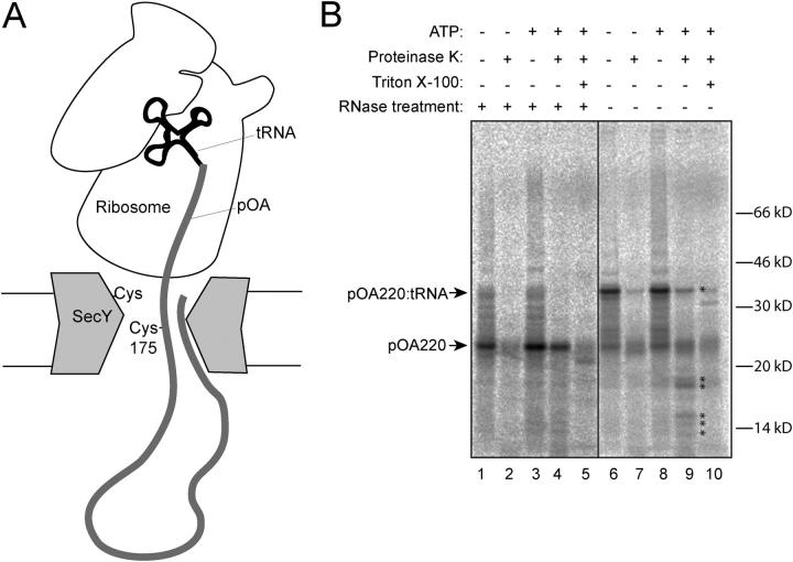
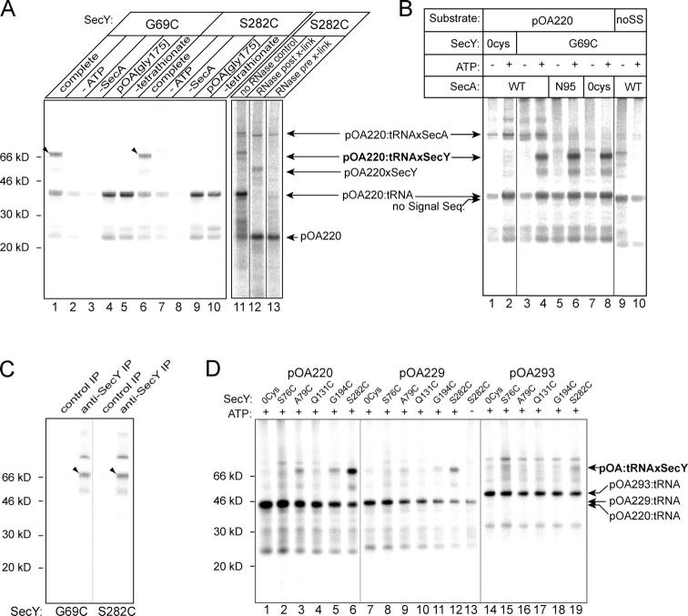
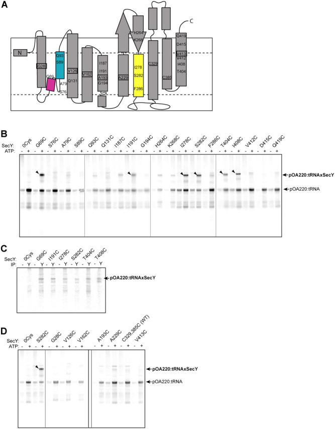
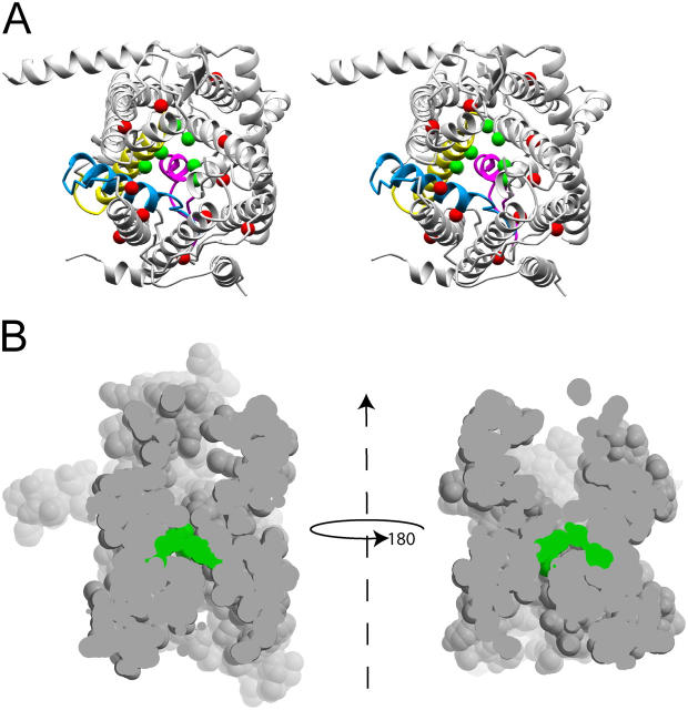
During their biosynthesis, many proteins pass through the membrane via a hydrophilic channel formed by the heterotrimeric Sec61/SecY complex. Whether this channel forms at the interface of multiple copies of Sec61/SecY or is intrinsic to a monomeric complex, as suggested by the recently solved X-ray structure of the Methanococcus jannaschii SecY complex, is a matter of contention. By introducing a single cysteine at various positions in Escherichia coli SecY and testing its ability to form a disulfide bond with a single cysteine in a translocating chain, we provide evidence that translocating polypeptides pass through the center of the SecY complex. The strongest cross-links were observed with residues that would form a constriction in an hourglass-shaped pore. This suggests that the channel makes only limited contact with a translocating polypeptide, thus minimizing the energy required for translocation.


Figures

Figure 1.
Figure 1.
Generating a translocation intermediate. (A) A tRNA-associated fragment of 35S-proOmpA (pOA) containing a single cysteine is trapped in the translocation channel. Another single cysteine is placed in SecY at a position that may line the channel. If the two cysteines are close to one another they can form an intermolecular disulfide bond upon exposure to oxidizing conditions. (B) A fragment of pOA containing 220 aa (pOA220) was synthesized in vitro by translation of truncated mRNA in the presence of [35S]methionine. Where indicated the samples were treated with RNase to remove the tRNA moiety. The translation products were then incubated with SecY complex–containing proteoliposomes and SecA in the presence or absence of ATP. Translocation was tested by treatment with protease in the absence or presence of Triton X-100. Asterisks highlight protected fragments of pOA220. Arrows point to positions of full-length pOA220 and full-length pOA220:tRNA.
Figure 2.
Figure 2.
Cysteines at positions 69 and 282 of SecY form a disulfide bond with pOA220:tRNA. (A) SecY mutants G69C and S282C were purified, reconstituted into proteoliposomes, incubated with cysteine-free SecA and 35S-pOA220:tRNA in the presence of ATP, and treated with 100 μM tetrathionate to facilitate disulfide bond formation. “Complete” contains all components; “−” indicates the omitted component. “pOA[Gly175]” is pOA220 with a glycine at position 175 instead of cysteine. Where indicated, samples were treated with RNase before or after cross-linking (pre- and post-X-link). Arrows point to the positions of the substrates and to cross-links with SecY and SecA. The cross-links to SecY are also highlighted by arrowheads. (B) Cross-linking was performed with pOA220 or substrate lacking the signal sequence (deletion of the NH2-terminal 21 aa of pOA220; noSS). SecY mutants lacked cysteines (0Cys) or contained a single cysteine at position 69 (G69C). SecA was either wild type (WT), or lacked three (N95) or all four (0Cys) cysteines. (C) Cross-linked samples were denatured and immunoprecipitated (IP) with affinity-purified polyclonal anti-SecY or control antibodies. (D) 35S-pOA:tRNA fragments of 220, 229, or 293 aa were translocated into proteoliposomes with SecY that contains no cysteines (0cys), or a single cysteine at positions 76, 79, 131, 194, or 282.
Figure 3.
Figure 3.
Identification of amino acids in SecY that contact the translocating polypeptide. (A) Cartoon showing the topology of SecY and positions where a cysteine was substituted for the endogenous residue. The two endogenous cysteines are underlined. Boxed numbers indicate positions that are predicted to face the exterior of the molecule. TM helices TM2a (magenta), TM2b (blue), and TM7 (yellow) are highlighted. (B) Purified SecY complexes with a single-cysteine substitution at a position predicted to face the interior of molecule were tested for their ability to form disulfide bonds with pOA220:tRNA in the presence of ATP as in Fig. 2; 0Cys, SecY lacking cysteines. Arrows indicate the substrate and its cross-link to SecY. Arrowheads highlight the strongest cross-links (>30% of total pOA220:tRNA linked to SecY). The gel shown is representative of at least three independent experiments. (C) Cross-linked samples were denatured and precipitated with affinity-purified polyclonal anti-SecY (Y) or control (−) antibodies. (D) SecY molecules with a single-cysteine substitution at positions on the exterior were screened for their ability to form disulfide bridges to pOA220. S282C is included as a positive control for cross-linking. 0Cys, SecY lacking cysteines; WT, wild-type SecY (cysteines at amino acids 329 and 385). An arrowhead highlights the cross-link to SecY S282C. The gel shown is representative of at least three independent experiments.
Figure 4.
Figure 4.
Positions in the X-ray structure of SecY that contact a translocating polypeptide. (A) Stereo view from the cytosol of the M. jannaschii SecY complex. Green spheres highlight the position of residues that in E. coli SecY give strong cross-links to a translocating chain (G69, I191, I278, S282, T404, I408). Red spheres indicate residues that did not give cross-links (G28, S76, S89, V126, Q131, V162, A193, A229, C329, C385, V413). TM helices TM2a (magenta), TM2b (blue), and TM7 (yellow) are highlighted. (B) Space-filling model of the SecY complex, viewed from the side. The complex has been sliced through the middle and opened to show the interior. Residues that form the strongest disulfide bonds with a translocation substrate are colored in green.

Similar articles

Cited by

References

    1. Bankaitis, V., B. Rasmussen, and P. Bassford. 1984. Intragenic suppressor mutations that restore export of maltose-binding protein with a truncated signal peptide. Cell. 37:243–252. - PubMed
    1. Beckmann, R., D. Bubeck, R. Grassucci, P. Penczek, A. Verschoor, G. Blobel, and J. Frank. 1997. Alignment of conduits for the nascent polypeptide chain in the ribosome-Sec61 complex. Science. 278:2123–2126. - PubMed
    1. Collinson, I., C. Breyton, F. Duong, C. Tziatzios, D. Schubert, E. Or, T. Rapoport, and W. Kuhlbrandt. 2001. Projection structure and oligomeric properties of a bacterial core protein translocase. EMBO J. 20:2462–2471. - PMC - PubMed
    1. Crowley, K.S., S.R. Liao, V.E. Worrell, G.D. Reinhart, and A.E. Johnson. 1994. Secretory proteins move through the endoplasmic reticulum membrane via an aqueous, gated pore. Cell. 78:461–471. - PubMed
    1. Duong, F. 2003. Binding, activation and dissociation of the dimeric SecA ATPase at the dimeric SecYEG translocase. EMBO J. 22:4375–4384. - PMC - PubMed

Publication types

MeSH terms