. 2005 Apr 25;169(2):233-8.
doi: 10.1083/jcb.200410027.

In vitro assembly and GTP hydrolysis by bacterial tubulins BtubA and BtubB

Affiliations

In vitro assembly and GTP hydrolysis by bacterial tubulins BtubA and BtubB

Christopher A Sontag et al. J Cell Biol. .

Abstract

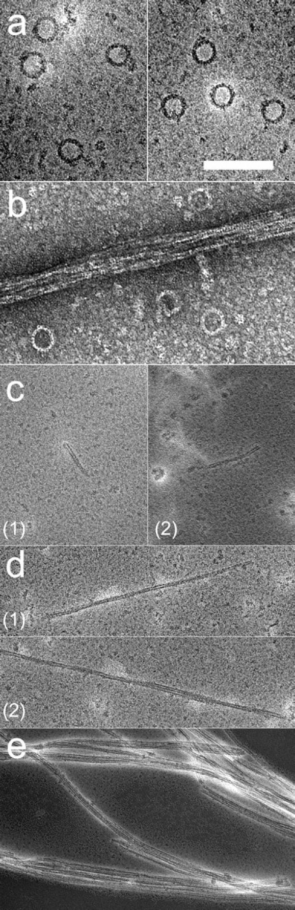
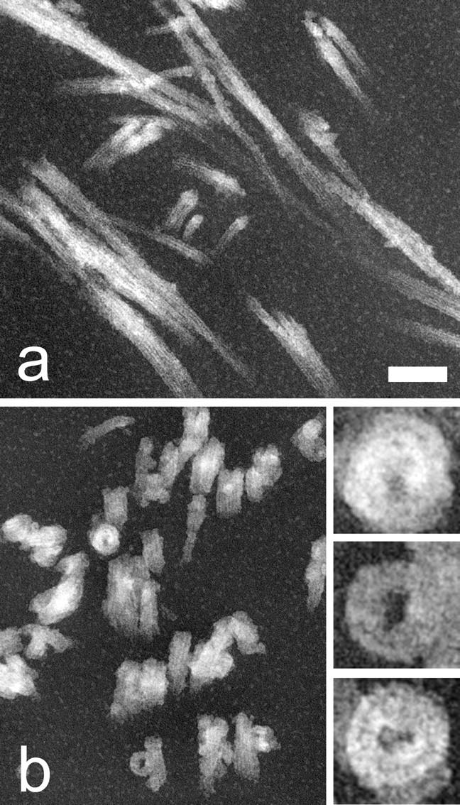
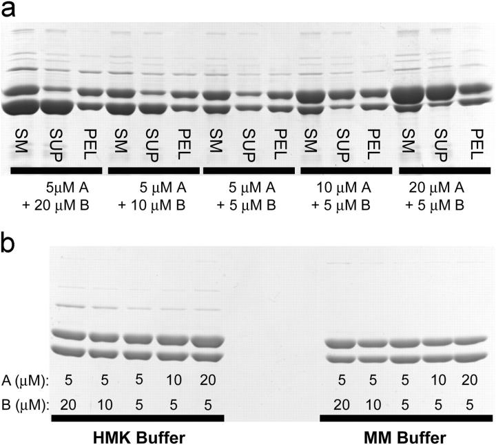
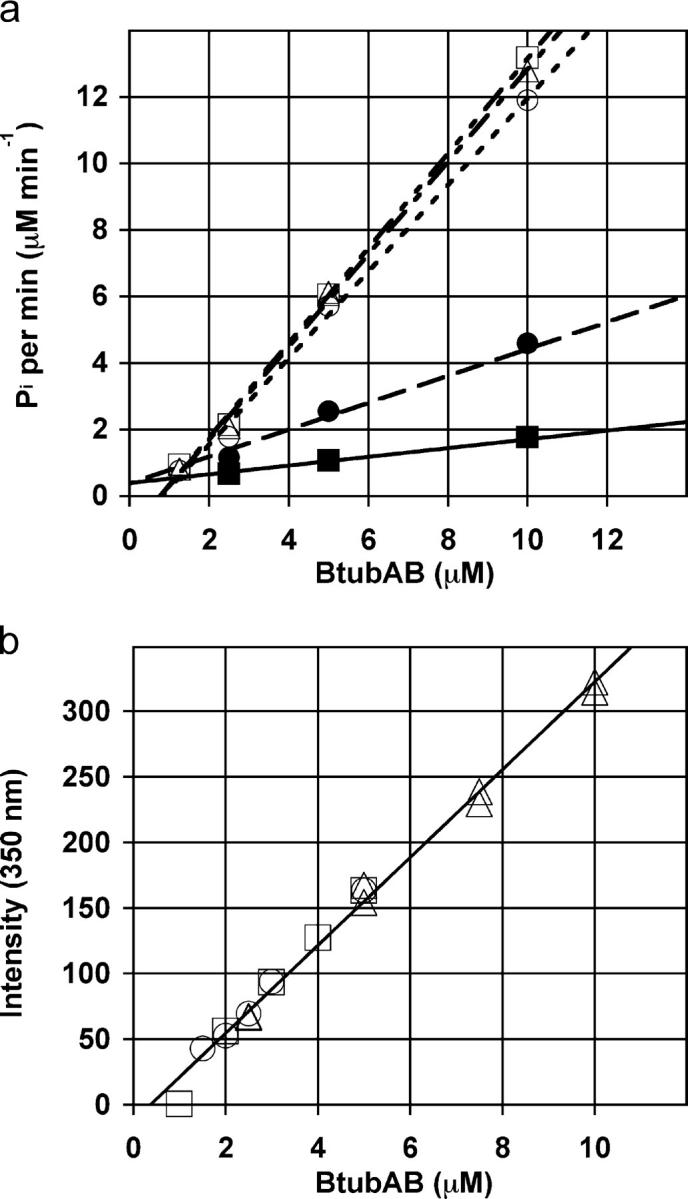
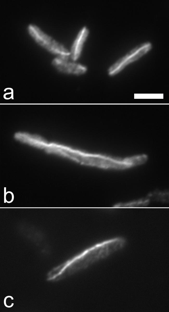
Arecent study identified genuine tubulin proteins, BtubA and BtubB, in the bacterial genus Prosthecobacter. We have expressed BtubA and BtubB in Escherichia coli and studied their in vitro assembly. BtubB by itself formed rings with an outer diameter of 35-36 nm in the presence of GTP or GDP. Mixtures of BtubB and BtubA formed long protofilament bundles, 4-7 protofilaments wide (20-30 protofilaments in the three-dimensional bundle). Regardless of the starting stoichiometry, the polymers always contained equal concentrations of BtubA and BtubB, suggesting that BtubA and B alternate along the protofilament. BtubA showed negligible GTP hydrolysis, whereas BtubB hydrolyzed 0.40 mol GTP per min per mol BtubB. This GTPase activity increased to 1.37 per min when mixed 1:1 with BtubA. A critical concentration of 0.4-1.0 microM was indicated by light scattering experiments and extrapolation of GTPase versus concentration, thus suggesting a cooperative assembly mechanism.


Figures

Figure 1.
Figure 1.
Negatively stained EM. (a) Rings formed by BtubB alone (5 μM). (b) A mixture of rings and protofilament bundles formed by BtubA + BtubB (5 μM each). Bar, 100 nm (for a and b). (c–e) Time-course EM for the polymerization of BtubA/B (1.2 μM each in HMK buffer) after addition of 200 μM GTP. (c) Short two-protofilament-wide bundles appear early in the reaction, 45 s. (d) At 2.5 min protofilament bundles are longer and add a third protofilament. (e) At 15 min protofilament bundles are longer and wider. Bar, 200 nm (for c–e).
Figure 2.
Figure 2.
Thin sections of BtubA/B polymers assembled in HMK buffer with 1 mM GTP. (a) A longitudinal section of protofilament bundles. (b) A cross section of protofilament bundles. Three bundles with an apparent lumen are shown in the three panels on the right. The bar represents 200 nm in a and b, and 25 nm in the three panels to the right of b.
Figure 3.
Figure 3.
Assembly of BtubA/B at various molar ratios in HMK buffer with 1 mM GTP, assayed by pelleting polymers. (a) Starting material (SM), supernatants (SUP), and resuspended pellets (PEL) were analyzed by SDS-PAGE. (b) The same reactions (as shown in panel a) plus reactions in MM buffer, but showing only the resuspended pellets for clarity. BtubA and BtubB are at a 1:1 molar ratio in all polymerized (pelleted) fractions.
Figure 4.
Figure 4.
GTPase activity and light-scattering studies of BtubA/B. (a) GTPase activity of BtubA/B in HMK buffer. The filled squares are BtubA; filled circles are BtubB; open squares, open circles, and open triangles represent three separate experiments with a 1:1 molar ratio of BtubA and BtubB. (b) BtubA and BtubB were mixed at 1:1 molar ratios in solution followed by the addition of 1 mM GTP at RT. Light-scattering data were collected at 350 nm and the plateau values were plotted versus the corresponding protein concentration. This plot represents the results from three separate experiments that were globally fit to a straight line. The line extrapolates to a critical concentration of 0.4 μM.
Figure 5.
Figure 5.
Immunofluorescent staining of BtubA/B structures in vivo. E. coli expressing both BtubA and BtubB were grown and fixed with a mixture of PFA and glutaraldehyde. Images are from cells that were stained with anti-BtubB antibody. Cells were grown at 37°C with 0.01% arabinose to mid-log phase. Bar, 2 μm.

References

    1. Addinall, S.G., E. Bi, and J. Lutkenhaus. 1996. FtsZ ring formation in fts mutants. J. Bacteriol. 178:3877–3784. - PMC - PubMed
    1. Anders, K.R., and D. Botstein. 2001. Dominant-lethal α-tubulin mutants defective in microtubule depolymerization in yeast. Mol. Biol. Cell. 12:3973–3986. - PMC - PubMed
    1. Bock, R.M., N.S. Ling, S.A. Morell, and S.H. Lipton. 1956. Ultraviolet absorption spectra of adenosine-5′-triphosphate and related 5′-ribonucleotides. Arch. Biochem. Biophys. 62:253–264. - PubMed
    1. Cayley, S., B.A. Lewis, H.J. Guttman, and M.T. Record Jr. 1991. Characterization of the cytoplasm of Escherichia coli K-12 as a function of external osmolarity: implications for protein-DNA interactions in vivo. J. Mol. Biol. 222:281–300. - PubMed
    1. Chen, Y., K. Bjornson, S.D. Redick, and H.P. Erickson. 2005. A rapid fluorescence assay for FtsZ assembly indicates cooperative assembly with a dimer nucleus. Biophys. J. 88:505–514. - PMC - PubMed

Publication types

MeSH terms