Comparative Study
. 2005 May 3;102(18):6437-42.
doi: 10.1073/pnas.0502467102. Epub 2005 Apr 25.

Distinct role of lymphocyte function-associated antigen-1 in mediating effective cytolytic activity by cytotoxic T lymphocytes

Affiliations
Comparative Study

Distinct role of lymphocyte function-associated antigen-1 in mediating effective cytolytic activity by cytotoxic T lymphocytes

Nadia Anikeeva et al. Proc Natl Acad Sci U S A. .

Abstract

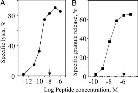
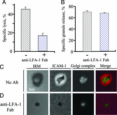
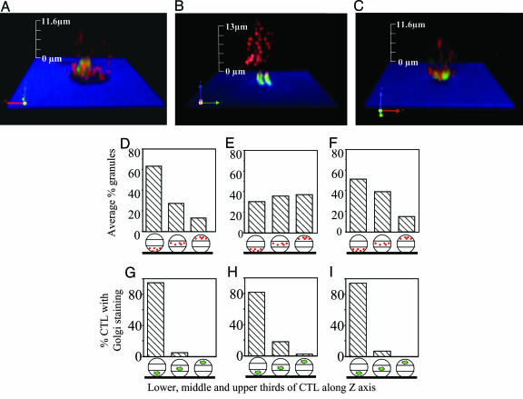
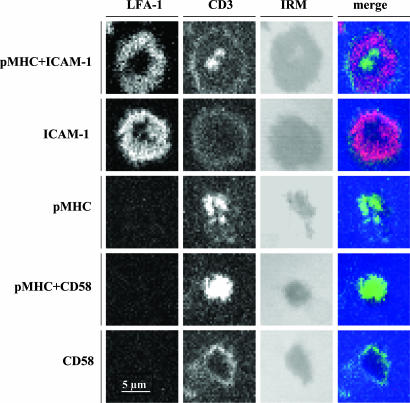
Lymphocyte function-associated antigen-1 (LFA-1) interaction with intercellular adhesion molecules (ICAMs) facilitates T cell antigen receptor (TCR)-mediated killing. To dissect TCR and LFA-1 contributions, we evaluated cytolytic activity and granule release by cytotoxic T lymphocytes (CTL) as well as intracellular granule redistribution and morphology of CTL stimulated with natural TCR ligand in the presence or absence of LFA-1 engagement. Although other adhesion mechanisms, e.g., CD2-CD58 interaction, could substitute for LFA-1 to trigger CTL degranulation, productive LFA-1 ligation was indispensable for effective target cell lysis by the released granules. LFA-1-mediated adhesion to glass-supported bilayers containing intercellular adhesion molecule-1 was characterized by a much larger junction area, marked by LFA-1 segregation, and a more compact cell shape compared with those observed for CD2-mediated adhesion to bilayers containing CD58. A larger contact induced by intercellular adhesion molecule 1 determined a unique positioning of granules near the interface. These data provide evidence that LFA-1 delivers a distinct signal essential for directing released cytolytic granules to the surface of antigen-bearing target cells to mediate the effective destruction of these cells by CTL.


Figures

Fig. 1.
Fig. 1.
Maximal granule release is not necessary for maximal target cell lysis. The concentration of GL9 peptide (arrow) required to achieve maximal specific lysis of HLA-A2+ target cells (JY) is ≈2 orders of magnitude lower than the peptide concentration necessary to induce maximal granule release by human Flu-specific CTL CER43. A representative experiment of five independent experiments is shown.
Fig. 2.
Fig. 2.
Inhibitory effect of anti-LFA-1 Fab on antigen-mediated cytolytic activity and the ability to form pSMAC by CTL. Inhibition of specific lysis of target cells (A) and release of cytolytic granules (B) by CTL at various peptide concentrations by Fab fragments of blocking TS1/22 anti-LFA-1 antibody are shown. Both assays were performed at effector-to-target cell ratio 1:1 and saturating peptide concentration (10-6 M) during 4 h. A representative result of five independent experiments is shown. Standard deviations are indicated. (C and D) TS1/22 anti-LFA-1 Fab blocks the formation of pSMAC (ICAM-1 ring) by CTL exposed to a glass-supported bilayer containing ICAM-1 and cognate pMHC. Images are taken at the level of the bilayer. The Golgi complex is recruited to the bilayer regardless of LFA-1-ICAM-1 interactions but to a different proximity. The Golgi complex was located 1.2 ± 0.4 and 1.9 ± 0.5 μm above the bilayer surface containing ICAM-1 + pMHC in the absence or presence of anti-LFA-1 Fab, respectively (P < 0.005). Merge image: ICAM-1 is red, and the Golgi complex is green.
Fig. 3.
Fig. 3.
Distribution of cytolytic granules and the Golgi complex in CTL exposed to various glass-supported bilayers. The distribution of the Golgi complex (green) and cytolytic granules (red) is recorded in various z sections (see Fig. 5) of CTL exposed to the bilayer containing either cognate pMHC and ICAM-1 (A), pMHC alone (B), or pMHC and CD58 (C); interference reflection microscopy indicates the level of bilayer (blue) and the CTL contact area (black). z sections were used to generate 3D images of Golgi complex (green) and cytolytic granule (red) intracellular distribution by using volocity software. Average percent of the granules (D-F) or Golgi complex (G-I) in the lower, middle, and upper thirds of all analyzed CTL exposed to bilayers containing ICAM-1 + pMHC (D and G), pMHC (E and H), or CD58 + pMHC (F and I) is shown.
Fig. 4.
Fig. 4.
Productive engagement of LFA-1 is required for its recruitment to the CTL contact surface and formation of the outer ring junction. CTL were exposed to various bilayers (indicated on the left), fixed, and stained with anti-LFA-1 mAb 24 (red), recognizing activated LFA-1 and anti-CD3 mAb (green). IRM shows the CTL contact area. LFA-1 ring staining is evident only on bilayers containing ICAM-1, regardless of the presence of cognate pMHC.

References

    1. Lowin, B., Hahne, M., Mattmann, C. & Tschopp, J. (1994) Nature 370, 650-652. - PubMed
    1. Berke, G. (1994) Annu. Rev. Immunol. 12, 735-773. - PubMed
    1. Tsomides, T. J., Aldovini, A., Johnson, R. P., Walker, B. D., Young, R. A. & Eisen, H. N. (1994) J. Exp. Med. 180, 1283-1293. - PMC - PubMed
    1. Sykulev, Y., Joo, M., Vturina, I., Tsomides, T. J. & Eisen, H. N. (1996) Immunity 4, 565-571. - PubMed
    1. Shaw, S., Luce, G. E., Quinones, R., Gress, R. E., Springer, T. A. & Sanders, M. E. (1986) Nature 323, 262-264. - PubMed

Publication types

MeSH terms

Substances

LinkOut - more resources