Comparative Study
. 2005 May 3;102(18):6332-7.
doi: 10.1073/pnas.0409142102. Epub 2005 Apr 25.

Evaluating putative mechanisms of the mitotic spindle checkpoint

Affiliations
Comparative Study

Evaluating putative mechanisms of the mitotic spindle checkpoint

Andreas Doncic et al. Proc Natl Acad Sci U S A. .

Abstract

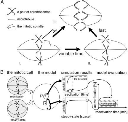
The mitotic spindle checkpoint halts the cell cycle until all chromosomes are attached to the mitotic spindles. Evidence suggests that the checkpoint prevents cell-cycle progression by inhibiting the activity of the APC-Cdc20 complex, but the precise mechanism underlying this inhibition is not yet known. Here, we use mathematical modeling to compare several mechanisms that could account for this inhibition. We describe the interplay between the capacities to strongly inhibit cell-cycle progression before spindle attachment on one hand and to rapidly resume cell-cycle progression once the last kinetochore is attached on the other hand. We find that inhibition that is restricted to the kinetochore region is not sufficient for supporting both requirements when realistic diffusion constants are considered. A mechanism that amplifies the checkpoint signal through autocatalyzed inhibition is also insufficient. In contrast, amplifying the signal through the release of a diffusible inhibitory complex can support reliable checkpoint function. Our results suggest that the design of the spindle checkpoint network is limited by physical constraints imposed by realistic diffusion constants and the relevant spatial and temporal dimensions where computation is performed.


Figures

Fig. 1.
Fig. 1.
Modeling framework. (A) A scheme of the mitotic spindle checkpoint. As long as even a single chromosome is unattached to the mitotic spindles, the anaphase does not commence, and the chromosomes do not separate (I). Once all chromosomes are properly attached, the cohesin connecting the chromosome is signaled for rapid degradation (II), and the anaphase starts (III). (B) To evaluate different models, we examined their compatibility with the physical parameters shown in Table 1. We focus on two properties of the checkpoint. First, we measure the extent of cell-cycle inhibition at steady state, before all chromosomes are attached. Second, we measure the time for reactivation once all kinetochores are properly attached. The mitotic cell is modeled as a sphere (radius R) with the last unattached kinetochore as a centrally located subsphere (radius ρ). At steady state, the extent of c-inhibition is measured by its fraction furthest away from the kinetochore. The reactivation time is defined as the time it takes for the level of uninhibited c proteins in the kinetochore vicinity to reach a certain threshold (taken here as 90%). A particular model with a given set of parameters defines a point in the “evaluation graph.” The working range corresponds to low enough inhibition (<5%) and rapid enough reactivation (<3 min).
Fig. 2.
Fig. 2.
Three possible models of the mitotic spindle checkpoint. (A) The direct inhibition model. Here the c proteins are only inhibited at the kinetochore itself. (B) The self-propagated inhibition model. The c proteins are inhibited at the kinetochore itself but also can catalyze the inhibition of additional c molecules everywhere in the nucleus at some rate κ.(C) The emitted inhibition model. Here the kinetochore catalyzes the formation of an inhibitory complex e*, which diffuses and inhibits the c molecules everywhere at some rate γ. The activated complexes also can decay spontaneously at some rate λ, and the total amount of e complexes is denoted as Etot. Note that the inhibited complex c* actually consists of both c and e. A scheme of each model is shown in A-C, and the corresponding equations are shown in D-F. G-L display an example of the steady-state activation level before the attachment and the temporal increase in activation once the kinetochore is attached for a typical set of parameters: Df = Di = 1 μm2·s-1, kinetochore size ρ = 0.01 μm, α = 0.02 s-1, κ = 0.38 μM-1·s-1, γ = 2.5 μM-1·s-1, Etot = 10Ctot, and λ = 0.1 s-1. Note that all concentration units are defined by the amount of c molecules, which is taken here as 1,000. Changing this number will somewhat change the levels of γ and κ, but it will have no effect on our qualitative results.
Fig. 3.
Fig. 3.
Evaluation graphs for the three models described in Fig. 2. The steady-state level of active c is plotted as a function of the reactivation time. Points were generated for increasing α, as indicated. The two dashed curved correspond to different diffusion constants of the free c (A and B) and the diffusion constant of the emitted inactive complex (C), as indicated. (A) The direct inhibition model. Here, α varies between ≈0.006 and ≈0.07 s-1.(B) Self-propagating inhibition model with κ = 0.05 μM-1·s-1 and α between ≈0.014 and ≈0.09 s-1. Increasing κ provides better inhibition but leads to infinite reactivation times. (C) The emitted inhibition model. Here, λ = 0.1 s-1, γ = 2 μM-1·s-1, Etot = 10Ctot and α between ≈0.007 and ≈0.14 s-1.

Similar articles

Cited by

References

    1. Hartwell, L. H. & Weinert, T. A. (1989) Science 246, 629-634. - PubMed
    1. Kastan, M. B. & Bartek, J. (2004) Nature 432, 316-323. - PubMed
    1. Hernando, E., Nahle, Z., Juan, G., Diaz-Rodriguez, E., Alaminos, M., Hemann, M., Michel, L., Mittal, V., Gerald, W., Benezra, R., et al. (2004) Nature 430, 797-802. - PubMed
    1. Rajagopalan, H. & Lengauer, C. (2004) Nature 432, 338-341. - PubMed
    1. Shonn, M. A., McCarroll, R. & Murray, A. W. (2000) Science 289, 300-303. - PubMed

Publication types

MeSH terms

Substances

LinkOut - more resources