

Brain mechanisms of involuntary visuospatial attention: an event-related potential study
- PMID: 15852465
- PMCID: PMC6871724
- DOI: 10.1002/hbm.20108
Brain mechanisms of involuntary visuospatial attention: an event-related potential study
Abstract
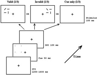
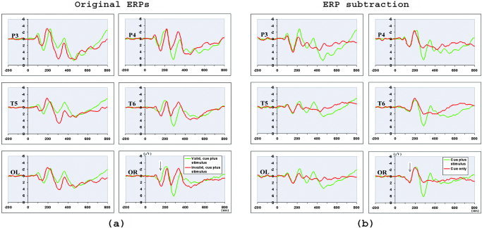
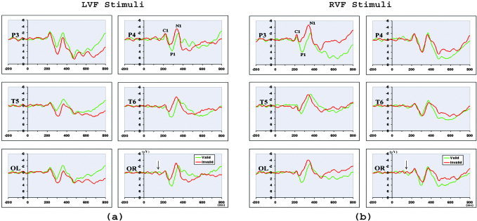
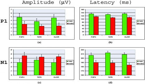
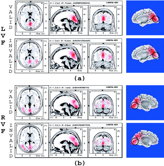
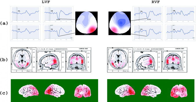
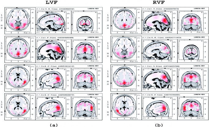
The brain mechanisms mediating visuospatial attention were investigated by recording event-related potentials (ERPs) during a line-orientation discrimination task. Nonpredictive peripheral cues were used to direct participant's attention involuntarily to a spatial location. The earliest attentional modulation was observed in the P1 component (peak latency about 130 ms), with the valid trials eliciting larger P1 than invalid trials. Moreover, the attentional modulations on both the amplitude and latency of the P1 and N1 components had a different pattern as compared to previous studies with voluntary attention tasks. In contrast, the earliest visual ERP component, C1 (peak latency about 80 ms), was not modulated by attention. Low-resolution brain electromagnetic tomography (LORETA) showed that the earliest attentional modulation occurred in extrastriate cortex (middle occipital gyrus, BA 19) but not in the primary visual cortex. Later attention-related reactivations in the primary visual cortex were found at about 110 ms after stimulus onset. The results suggest that involuntary as well as voluntary attention modulates visual processing at the level of extrastriate cortex; however, at least some different processes are involved by involuntary attention compared to voluntary attention. In addition, the possible feedback from higher visual cortex to the primary visual cortex is faster and occurs earlier in involuntary relative to voluntary attention task.
Figures
Similar articles
-
The attentional effects of peripheral cueing as revealed by two event-related potential studies.Clin Neurophysiol. 2001 Jan;112(1):172-85. doi: 10.1016/s1388-2457(00)00500-9. Clin Neurophysiol. 2001. PMID: 11137676 Clinical Trial.
-
Event-related potentials reveal dissociable mechanisms for orienting and focusing visuospatial attention.Brain Res Cogn Brain Res. 2005 May;23(2-3):341-53. doi: 10.1016/j.cogbrainres.2004.11.014. Brain Res Cogn Brain Res. 2005. PMID: 15820641 Free PMC article.
-
Delayed striate cortical activation during spatial attention.Neuron. 2002 Aug 1;35(3):575-87. doi: 10.1016/s0896-6273(02)00781-x. Neuron. 2002. PMID: 12165478
-
Dynamics of emotional effects on spatial attention in the human visual cortex.Prog Brain Res. 2006;156:67-91. doi: 10.1016/S0079-6123(06)56004-2. Prog Brain Res. 2006. PMID: 17015075 Review.
-
A new neural framework for visuospatial processing.Nat Rev Neurosci. 2011 Apr;12(4):217-30. doi: 10.1038/nrn3008. Nat Rev Neurosci. 2011. PMID: 21415848 Free PMC article. Review.
Cited by
-
Backward masked fearful faces enhance contralateral occipital cortical activity for visual targets within the spotlight of attention.Soc Cogn Affect Neurosci. 2011 Oct;6(5):639-45. doi: 10.1093/scan/nsq076. Epub 2010 Aug 11. Soc Cogn Affect Neurosci. 2011. PMID: 20702500 Free PMC article.
-
Event-related brain potentials in the study of inhibition: cognitive control, source localization and age-related modulations.Neuropsychol Rev. 2014 Dec;24(4):461-90. doi: 10.1007/s11065-014-9275-4. Epub 2014 Nov 19. Neuropsychol Rev. 2014. PMID: 25407470 Review.
-
Nicotine modulation of information processing is not limited to input (attention) but extends to output (intention).Psychopharmacology (Berl). 2010 May;209(4):291-302. doi: 10.1007/s00213-010-1788-9. Epub 2010 Mar 23. Psychopharmacology (Berl). 2010. PMID: 20309531 Free PMC article. Clinical Trial.
-
Magnocellular and parvocellular influences on reflexive attention.Vision Res. 2011 Aug 15;51(16):1820-8. doi: 10.1016/j.visres.2011.06.012. Epub 2011 Jun 23. Vision Res. 2011. PMID: 21723311 Free PMC article.
-
Spatiotemporal brain mapping of spatial attention effects on pattern-reversal ERPs.Hum Brain Mapp. 2012 Jun;33(6):1334-51. doi: 10.1002/hbm.21285. Epub 2011 Apr 15. Hum Brain Mapp. 2012. PMID: 21500317 Free PMC article.
References
-
- Aine CJ, Supek S, George JS (1995): Temporal dynamics of visual‐evoked neuromagnetic sources: effects of stimulus parameters and selective attention. Int J Neurosci 80: 79–104. - PubMed
-
- Aine CJ, Supek S, George JS, Ranken D, Lewine J, Sanders J, Best E, Tiee W, Flynn ER, Wood CC (1996): Retinotopic organization of human visual cortex: departures from the classical model. Cereb Cortex 6: 354–361. - PubMed
-
- Anllo‐Vento L (1995): Shifting attention in visual space: the effects of peripheral cueing on brain cortical potentials. Int J Neurosci 80: 353–370. - PubMed
-
- Anllo‐Vento L, Hillyard SA (1996): Selective attention to the color and direction of moving stimuli: electrophysiological correlates of hierarchical feature selection. Percep Psychophys 58: 191–206. - PubMed
Publication types
MeSH terms
Grants and funding
LinkOut - more resources
Full Text Sources
