

The effect of social content on deductive reasoning: an fMRI study
- PMID: 15852469
- PMCID: PMC6871752
- DOI: 10.1002/hbm.20114
The effect of social content on deductive reasoning: an fMRI study
Abstract
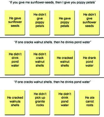
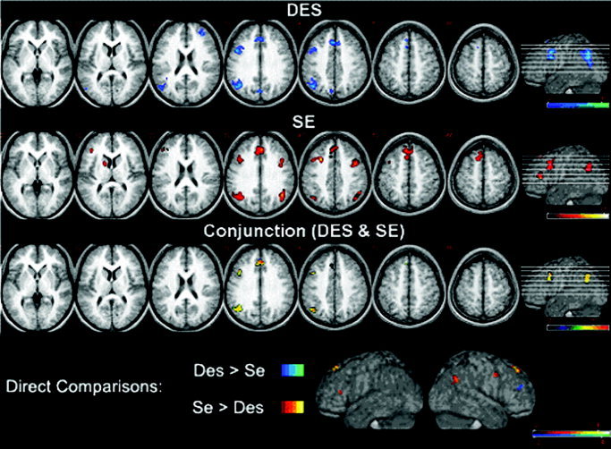
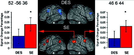
Psychological studies of deductive reasoning have shown that subjects' performance is affected significantly by the content of the presented stimuli. Specifically, subjects find it easier to reason about contexts and situations with a social content. In the present study, the effect of content on brain activation was investigated with functional magnetic resonance imaging (fMRI) while subjects were solving two versions of the Wason selection task, which previous behavioral studies have shown to elicit a significant content effect. One version described an arbitrary relation between two actions (Descriptive: "If someone does ..., then he does ..."), whereas the other described an exchange of goods between two persons (Social-Exchange: "If you give me ..., then I give you ..."). Random-effect statistical analyses showed that compared to baseline, both tasks activated frontal medial cortex and left dorsolateral frontal and parietal regions, confirming the major role of the left hemisphere in deductive reasoning. In addition, although the two reasoning conditions were identical in logical form, the social-exchange task was also associated with right frontal and parietal activations, mirroring the left-sided activations common to both reasoning tasks. These results suggest that the recruitment of the right hemisphere is dependent on the content of the stimuli presented.
Figures
References
-
- Acuna BD, Eliassen JC, Donoghue JP, Sanes JN (2002): Frontal and parietal lobe activation during transitive inference in humans. Cereb Cortex 12: 1312–1321. - PubMed
-
- Adolphs R (2001): The neurobiology of social cognition. Curr Opin Neurobiol 11: 231–239. - PubMed
-
- Adolphs R (2003): Cognitive neuroscience of human social behaviour. Nat Rev Neurosci 4: 165–178. - PubMed
-
- Alexander GE, DeLong MR, Strick PL (1986): Parallel organization of functionally segregated circuits linking basal ganglia and cortex. Annu Rev Neurosci 9: 357–381. - PubMed
Publication types
MeSH terms
LinkOut - more resources
Full Text Sources
