. 2005 May;166(5):1309-20.
doi: 10.1016/S0002-9440(10)62350-4.

In response to protein load podocytes reorganize cytoskeleton and modulate endothelin-1 gene: implication for permselective dysfunction of chronic nephropathies

Affiliations

In response to protein load podocytes reorganize cytoskeleton and modulate endothelin-1 gene: implication for permselective dysfunction of chronic nephropathies

Marina Morigi et al. Am J Pathol. 2005 May.

Abstract

Effacement of podocyte foot processes occurs in many proteinuric nephropathies and is accompanied by rearrangement of the actin cytoskeleton. Here, we studied whether protein overload affects intracellular pathways, leading to cytoskeletal architecture changes and ultimately to podocyte dysfunction. Mouse podocytes bound and endocytosed both albumin and IgG via receptor-specific mechanisms. Protein overload caused redistribution of F-actin fibers instrumental to up-regulation of the prepro-endothelin (ET)-1 gene and production of the corresponding peptide. Increased DNA-binding activity for nuclear factor (NF)-kappaB and Ap-1 nuclear proteins was measured in nuclear extracts of podocytes exposed to excess proteins. Both Y27632, which inhibits Rho kinase-dependent stress fiber formation, and jasplakinolide, an F-actin stabilizer, decreased NF-kappaB and Ap-1 activity and reduced ET-1 expression. This suggested a role for the cytoskeleton, through activated Rho, in the regulation of the ET-1 peptide. Focal adhesion kinase (FAK), an integrin-associated nonreceptor tyrosine kinase, was phosphorylated by albumin treatment via Rho kinase-triggered actin reorganization. FAK activation led to NF-kappaB- and Ap-1-dependent ET-1 expression. These data suggest that reorganization of the actin cytoskeletal network in response to protein load is implicated in modulation of the ET-1 gene via Rho kinase-dependent FAK activation of NF-kappaB and Ap-1 in differentiated podocytes. Increased ET-1 generation might alter glomerular permselectivity and amplify the noxious effect of protein overload on dysfunctional podocytes.


Figures
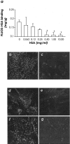
Figure 1
Figure 1
Binding and uptake of HSA and human IgG on differentiated podocytes. Top: Binding studies. a: Confluent differentiated podocytes were incubated with 50 μg/ml of FLUOS-HSA with or without increasing concentrations of cold HSA. Complete inhibition of FLUOS-HSA binding by unlabeled HSA showed that binding was specific and not due to an interaction of FLUOS-HSA with plasma membrane (n = 5 experiments). Data are expressed as mean ± SE. *, P < 0.05 versus 0 mg/ml HSA. Representative images of binding of 50 μg/ml of FITC-IgG on podocytes in the absence (b) or presence of 5 mg/ml of cold IgG (c). Bottom: Uptake studies. Cellular staining of 50 μg/ml of FLUOS-HSA incubated without (d) or with (e) 5 mg/ml of unlabeled HSA. Endocytosis of 50 μg/ml of FITC-IgG in podocytes in the absence (f) or presence (g) of 5 mg/ml of unlabeled IgG. Original magnifications, ×600.
Figure 2
Figure 2
Reorganization of F-actin cytoskeleton in podocytes exposed to protein overload. Confluent differentiated podocytes exhibited a pattern of F-actin filaments distributed as stress fiber-like bundles along the axis of the cells after 6 hours (a) incubation with medium alone. Redistribution of F-actin fibers to the periphery of the cells was observed in podocytes exposed to HSA already at 30 minutes (b) that was maintained at 1 (c), 2 (d), 6 (e), and 24 hours (f). A similar effect was observed after podocyte exposure to IgG for 6 and 24 hours (g, h). Original magnifications, ×600.
Figure 3
Figure 3
Double immunolabeling for F-actin and ZO-1 in podocytes challenged with HSA. Immunofluorescence images of F-actin and ZO-1 in podocytes exposed to medium alone (a) or HSA for 6 hours (b) indicate that protein overload rearranged F-actin fibers at the periphery of the cells as outlined by ZO-1 staining (b). Original magnifications, ×1000.
Figure 4
Figure 4
Expression of synaptopodin in podocytes exposed to HSA or IgG. a: High level of synaptopodin in a punctate pattern was observed along actin filament in unstimulated cells. After 6 hours of exposure of podocytes with HSA (b) or IgG (c) at the concentration of 10 mg/ml, synaptopodin was markedly reduced in respect to control cells. Original magnifications, ×600.
Figure 5
Figure 5
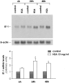
ET-1 mRNA expression in podocytes exposed to protein overload. Top: Northern blot experiments were performed using total RNA isolated from podocytes exposed to medium alone (control) or albumin (10 mg/ml) for 6, 24, and 48 hours. The results are representative of four independent experiments. Bottom: Densitometric analysis of autoradiograph signals for ET-1. The optical density of the autoradiographic signals was quantified and calculated as the ratio of ET-1 to β-actin mRNA. Results (mean ± SE) are expressed as fold increase greater than control (considered as 1) in densitometric arbitrary units. °, P < 0.01 versus control.
Figure 6
Figure 6
Effect of the cytoskeleton inhibitors Y27632 and jasplakinolide on albumin-induced ET-1 gene expression. Cells were treated with medium alone or with HSA (10 mg/ml) for 3, 6, 15, and 24 hours in the presence or absence of Y27632 (10 μmol/L), an inhibitor of Rho kinase pathway, or jasplakinolide (200 nmol/L), an F-actin stabilizer. ET-1 mRNA was assessed by real-time PCR. The results shown are mean ± SE of five independent experiments. °, P < 0.01 versus control; *, P < 0.05; **, P < 0.01 versus HSA.
Figure 7
Figure 7
ET-1 production in podocytes exposed to albumin and effect of Y27632 and jasplakinolide. Podocytes were incubated with medium alone or with HSA (10 mg/ml) for 3, 6, 15, and 24 hours in the presence or absence of Y27632 (10 μmol/L) and jasplakinolide (200 nmol/L). ET-1 production was measured in cell supernatants by RIA. Data are expressed as mean ± SE. °, P < 0.01 versus control; *, P < 0.05; **, P < 0.01 versus HSA; #, P < 0.01 versus HSA + Y27632.
Figure 8
Figure 8
Activation of NF-κB and Ap-1 in protein-laden podocytes. NF-κB and AP-1 activity was evaluated by EMSA in nuclear extracts from podocytes exposed for 30 minutes to medium alone, HSA, or IgG (10 mg/ml). To demonstrate the specificity of binding of the NF-κB and Ap-1 oligonucleotides, a 100-fold molar excess unlabeled (cold) nucleotides were used to compete with the labeled NF-κB or AP-1 probes for binding to nuclear proteins. Results are representative of three experiments.
Figure 9
Figure 9
Effect of Y27632 and jasplakinolide on NF-κB and Ap-1 activation induced by protein overload. Top: EMSA for NF-κB and Ap-1 was performed in nuclear extracts from podocytes exposed for 30 minutes to medium alone or HSA (10 mg/ml), in the presence or absence of Y27632 (10 μmol/L) and jasplakinolide (200 nmol/L). The results are representative of five independent experiments using different nuclear extracts. Bottom: Densitometric analysis of autoradiographic signals of NF-κB and AP-1. Results are mean ± SE. °, P < 0.01 versus control; *, P < 0.05; and **, P < 0.01 versus HSA.
Figure 10
Figure 10
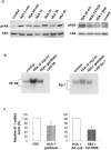
Role of FAK on ET-1 gene activation/expression in HSA-treated podocytes. a: Left: Activation of FAK in podocytes exposed to HSA (10 mg/ml) for 5 minutes, 30 minutes, and 1, 2, 3, and 6 hours. a: Right: Effect of Y27632 (10 μmol/L) and jasplakinolide (jasp, 200 nmol/L) on FAK phosphorylation in podocytes treated for 30 minutes with HSA. Cell lysates were analyzed by Western blot using antibody against the phosphorylated form. The blots were stripped and reprobed with an antibody anti-nonphosphorylated FAK to confirm equal loading of the proteins on the gel. The blot is representative of three independent experiments. b: Effect of genistein on NF-κB and Ap-1 activation induced by protein overload. EMSA for NF-κB and Ap-1 was performed in nuclear extracts from podocytes exposed for 30 minutes to medium alone or HSA in the presence or absence of genistein (25 μmol/L). The autoradiographs are representative of three independent experiments using different nuclear extracts. c: Effect of inhibition of FAK phosphorylation on ET-1 expression. Left: Cells were treated with medium alone or with HSA for 3 hours in the presence or absence of genistein. Right: Cells were transfected with a replication-defective adenovirus encoding FAK-related nonkinase (Ad-FRNK), an endogenous inhibitor of FAK activity or Ad-null, before HSA exposure. ET-1 mRNA was assessed by real-time PCR. *, P < 0.01 versus HSA + Ad-null.
Figure 11
Figure 11
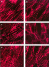
Effect of ET-1 on cytoskeletal F-actin distribution in podocytes. Immunofluorescence staining of F-actin fibers in podocytes exposed for 2, 6, and 15 hours to control medium (a, c, e) or ET-1 (b, d, f). In unstimulated cells F-actin microfilaments were arranged in parallel, whereas on exposure to ET-1 (100 nmol/L) for 2 and 6 hours, this pattern changed leading to F-actin redistribution at the cell periphery. This effect partially recovered at 15 hours. Original magnifications, ×600.
Figure 12
Figure 12
Proposed pathway mediating protein overload-induced ET-1 expression. Ultrafiltered plasma proteins, by binding to specific receptors on podocyte surface, cause cytoskeleton rearrangement and new F-actin stress fiber formation via Rho kinase pathway. Rho kinase-triggered actin reorganization leads to FAK phosphorylation. FAK activation integrates signals from cytoskeleton to the downstream activation of NF-κB and Ap-1 which in turn translocate to the nucleus and promote pre-pro ET-1 gene transcription.

References

    1. Olson JL, Hostetter TH, Rennke HG, Brenner BM, Venkatachalam MA. Altered glomerular permselectivity and progressive sclerosis following extreme ablation of renal mass. Kidney Int. 1982;22:112–126. - PubMed
    1. Brenner BM, Meyer TW, Hostetter TH. Dietary protein intake and progressive nature of kidney disease: the role of hemodynamically mediated glomerular injury in the pathogenesis of progressive glomerular sclerosis in aging, renal ablation, and intrinsic renal disease. N Engl J Med. 1982;307:652–659. - PubMed
    1. Anderson SMT, Rennke HG, Brenner BM. Control of glomerular hypertension limits glomerular injury in rats with reduced renal mass. J Clin Invest. 1985;76:612–619. - PMC - PubMed
    1. Fries JW, Sandstrom DJ, Meyer TW, Rennke HG. Glomerular hypertrophy and epithelial cell injury modulate progressive glomerulosclerosis in the rat. Lab Invest. 1989;60:205–218. - PubMed
    1. Remuzzi G, Bertani T. Is glomerulosclerosis a consequence of altered glomerular permeability to macromolecules? Kidney Int. 1990;38:384–394. - PubMed

Publication types

MeSH terms

LinkOut - more resources