. 2005 May;166(5):1321-32.
doi: 10.1016/s0002-9440(10)62351-6.

Induction of epithelial-mesenchymal transition in alveolar epithelial cells by transforming growth factor-beta1: potential role in idiopathic pulmonary fibrosis

Affiliations

Induction of epithelial-mesenchymal transition in alveolar epithelial cells by transforming growth factor-beta1: potential role in idiopathic pulmonary fibrosis

Brigham C Willis et al. Am J Pathol. 2005 May.

Abstract

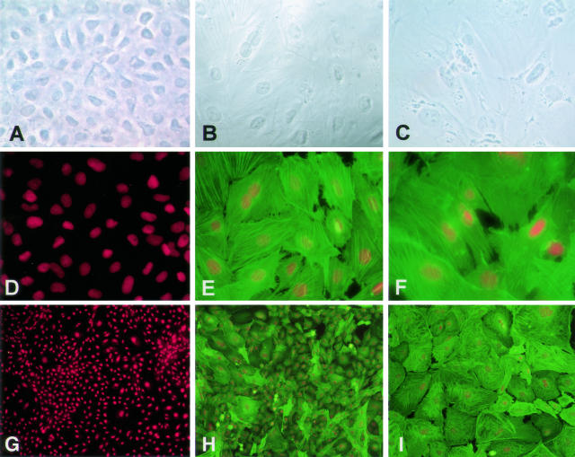
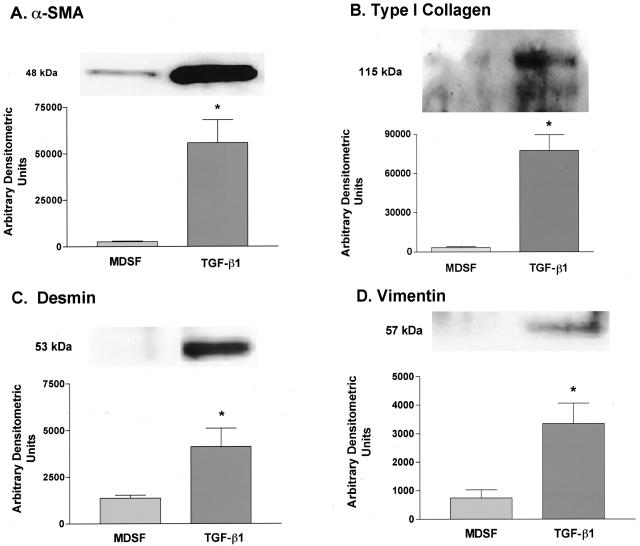
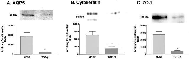
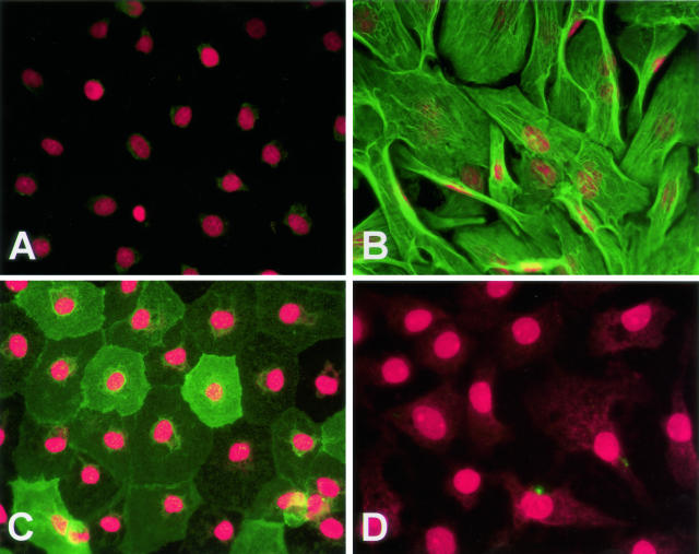
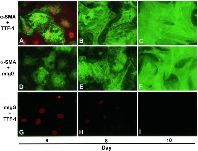
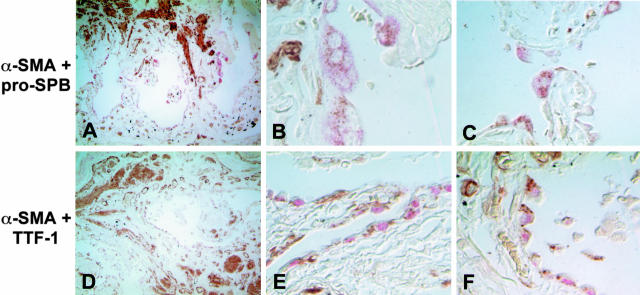
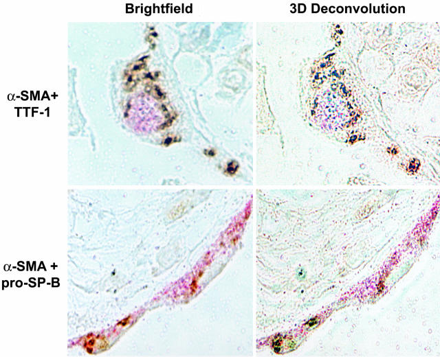
The hallmark of idiopathic pulmonary fibrosis (IPF) is the myofibroblast, the cellular origin of which in the lung is unknown. We hypothesized that alveolar epithelial cells (AECs) may serve as a source of myofibroblasts through epithelial-mesenchymal transition (EMT). Effects of chronic exposure to transforming growth factor (TGF)-beta1 on the phenotype of isolated rat AECs in primary culture and a rat type II cell line (RLE-6TN) were evaluated. Additionally, tissue samples from patients with IPF were evaluated for cells co-expressing epithelial (thyroid transcription factor (TTF)-1 and pro-surfactant protein-B (pro-SP-B), and mesenchymal (alpha-smooth muscle actin (alpha-SMA)) markers. RLE-6TN cells exposed to TGF-beta1 for 6 days demonstrated increased expression of mesenchymal cell markers and a fibroblast-like morphology, an effect augmented by tumor necrosis factor-alpha (TNF-alpha). Exposure of rat AECs to TGF-beta1 (100 pmol/L) resulted in increased expression of alpha-SMA, type I collagen, vimentin, and desmin, with concurrent transition to a fibroblast-like morphology and decreased expression of TTF-1, aquaporin-5 (AQP5), zonula occludens-1 (ZO-1), and cytokeratins. Cells co-expressing epithelial markers and alpha-SMA were abundant in lung tissue from IPF patients. These results suggest that AECs undergo EMT when chronically exposed to TGF-beta1, raising the possibility that epithelial cells may serve as a novel source of myofibroblasts in IPF.


Figures

Figure 1
Figure 1
Effects of TGF-β1 ± TNF-α on cell morphology and expression of α-SMA in RLE-6TN cells. Immunoreactivity for α-SMA (green) was assessed by immunofluorescence on day 6, and accompanying phase images of cell morphology were obtained. Expression of α-SMA and assumption of a fibroblast-like morphology was induced in monolayers exposed to TGF-β1 and was seen in 100% of cells in cultures treated with TGF-β1 and TNF-α. A: Phase image of monolayer in media only. B: Phase image of monolayer in media + TGF-β1. C: Phase image of monolayer in media + TGF-β1 + TNF-α. D and G: Monolayer in media only reacted with anti-α-SMA mAb. E and H: Monolayer in media + TGF-β1 reacted with anti-α-SMA mAb. F and I: Monolayer in media + TGF-β1 + TNF-α reacted with anti-α-SMA mAb. Red staining represents propidium iodide-stained nuclei. Photographs are representative of >12 cultures from more than three separate experiments. Original magnification, ×400 for A through F; ×100 for G through I.
Figure 2
Figure 2
Effects of TGF-β1 on expression of mesenchymal cell markers in AECs. Representative Western blots (n = 4 for α-SMA; n = 3 for type I collagen, vimentin, and desmin) and densitometric analyses demonstrate increases in expression of α-SMA, type I collagen, vimentin, and desmin on day 14 in AECs treated with TGF-β1 for 12 days. eIF was used as a loading control for all blots, and densitometric analyses were adjusted to account for relative differences in eIF intensity (data not shown). A: α-SMA expression increased 21-fold over untreated cultures on day 14 in treated AECs. B: Type I collagen expression increased 27-fold over untreated cultures on day 14 in treated AECs. C: Desmin expression increased threefold over untreated cultures on day 14 in treated AECs. D: Vimentin expression increased 4.5-fold over untreated cultures on day 14 in treated AECs. *Significantly different from MDSF.
Figure 3
Figure 3
Effects of TGF-β1 on AEC expression of epithelial cell markers. Representative Western blots (n = 4 for AQP5, n = 3 for pancytokeratin and ZO-1) and densitometric analyses demonstrate decreases in expression of AQP5, pancytokeratin, and ZO-1 on day 14 in AECs treated with TGF-β1 for 12 days. eIF was used as a loading control for all blots, and densitometric analyses were adjusted to account for relative differences in eIF intensity (data not shown). Expression of AQP5 (A), cytokeratins (B), and ZO-1 (C) decreased to about 10, 29, and 6% of untreated levels, respectively. *Significantly different from MDSF.
Figure 4
Figure 4
Effects of TGF-β1 on AEC morphology and associated expression of α-SMA and AQP5. Immunoreactivity for α-SMA (green, A and B) and AQP5 (green, C and D) was assessed by immunofluorescence on day 14. Expression of α-SMA was undetectable in untreated monolayers but was strongly expressed in a fibril-associated pattern in TGF-β1-treated cultures. AQP5 was highly expressed in untreated cultures but was undetectable in monolayers exposed to TGF-β1. A: Monolayer in MDSF reacted with anti-α-SMA mAb. B: Monolayer in MDSF + TGF-β1 reacted with anti-α-SMA mAb. C: Monolayer in MDSF reacted with anti-AQP5 polyclonal Ab. D: Monolayer in MDSF + TGF-β1 reacted with anti-AQP5 polyclonal Ab. Control slides using mIgG revealed no significant staining (not shown). Red staining represents propidium iodide-stained nuclei. Photographs are representative of >12 monolayers from more than five separate experiments. Original magnification, ×400.
Figure 5
Figure 5
Colocalization of α-SMA and TTF-1 in primary AECs during epithelial-mesenchymal transition. Immunoreactivity for α-SMA (green) and TTF-1 (red) was assessed on days 6, 8, and 10 of primary culture of AECs in MDSF + TGF-β1 + TNF-α. On day 6, individual AECs were identified that co-express nuclear TTF-1 and cytoplasmic α-SMA. Expression of α-SMA increased gradually over time in culture (A to C, D to F) and paralleled a concomitant decrease in expression of TTF-1 (A to C, G to I) along with a transition from an epithelial to a fibroblast-like morphology. Photographs are representative of >12 monolayers from more than three separate experiments. Original magnification, ×600.
Figure 6
Figure 6
Co-expression of myofibroblast and AEC markers in AECs of lung tissue from humans with IPF. Low-power images (original magnification, ×100) reveal severe destruction of normal lung architecture due to fibrosis (A and D). Pink color represents Fast Red staining of SP-B (A to C) or TTF-1 (D to F). Brown color represents diaminobenzidine staining of α-SMA. High-power images (original magnification, ×400; two representative fields from within the low-power field shown) reveal abundant epithelial cells adjacent to fibroblastic foci co-expressing SP-B (pink, cytoplasmic) and α-SMA (brown, cytoplasmic) (B and C) or TTF-1 (pink, nuclear) and α-SMA (brown, cytoplasmic) (E and F). Colocalization of both markers was seen in all three patients examined.
Figure 7
Figure 7
Colocalization of myofibroblast and AEC markers in AECs of lung tissue from humans with IPF using three-dimensional deconvolution microscopy. High-power images (original magnification, ×100) of single epithelial cells were taken at sequential 0.1-μm z axis depths >3 μm. Resultant image stacks were analyzed using a three-dimensional deconvolution algorithm. Pro-SPB and TTF-1 (pink) colocalized with α-SMA (brown) to the same optical section in all cells analyzed.

References

    1. Crystal RG, Bitterman PB, Mossman B, Schwarz MI, Sheppard D, Almasy L, Chapman HA, Friedman SL, King TE, Jr, Leinwand LA, Liotta L, Martin GR, Schwartz DM, Schultz GS, Wagner CR, Musson RA. Future research directions in idiopathic pulmonary fibrosis. Am J Respir Cell Mol Biol. 2002;166:236–246. - PubMed
    1. Travis WD, King TE, Jr, Bateman ED, Lynch DA. American Thoracic Society/European Respiratory Society international multidisciplinary consensus classification of the idiopathic interstitial pneumonias. Am J Resp Crit Care Med. 2002;265:277–304. - PubMed
    1. Selman M, Pardo A. The epithelial/fibroblastic pathway in the pathogenesis of idiopathic pulmonary fibrosis. Am J Respir Cell Mol Biol. 2003;29:S93–S96. - PubMed
    1. Chilosi M, Poletti V, Zamo A, Lestani M, Montagna L, Piccoli P, Pedron S, Bertaso M, Scarpa A, Murer B, Cancellieri A, Maestro R, Semenzato G, Doglioni C. Aberrant Wnt/β-catenin pathway activation in idiopathic pulmonary fibrosis. Am J Pathol. 2003;162:1495–1502. - PMC - PubMed
    1. Pittet JF, Griffiths MJD, Geiser T, Kaminski N, Dalton SL, Huang X, Brown LAS, Gotwals PJ, Koteliansky VE, Matthay MA, Sheppard D. TGF-β is a critical mediator of acute lung injury. J Clin Invest. 2001;107:1537–1544. - PMC - PubMed

Publication types

MeSH terms