. 2005 May;166(5):1353-65.
doi: 10.1016/S0002-9440(10)62354-1.

Absence of proteinase-activated receptor-1 signaling affords protection from bleomycin-induced lung inflammation and fibrosis

Affiliations

Absence of proteinase-activated receptor-1 signaling affords protection from bleomycin-induced lung inflammation and fibrosis

David C J Howell et al. Am J Pathol. 2005 May.

Abstract

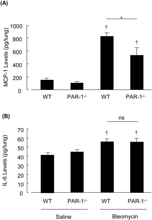
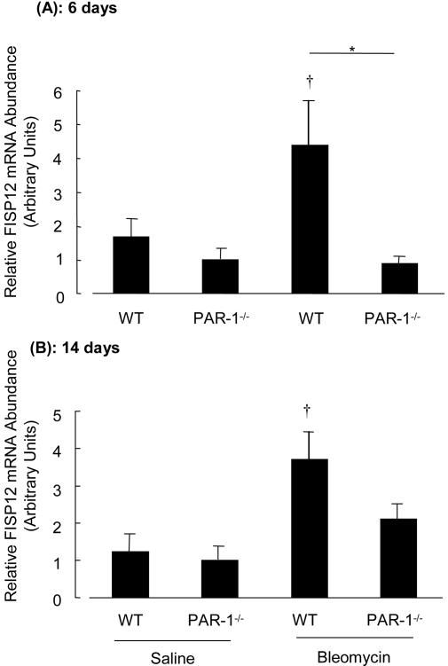
Activation of the coagulation cascade is commonly observed in the lungs of patients with both acute and chronic inflammatory and fibrotic lung disorders, as well as in animal models of these disorders. The aim of this study was to examine the contribution of the major thrombin receptor, proteinase-activated receptor-1 (PAR-1), during the acute inflammatory and chronic fibrotic phases of lung injury induced by intratracheal instillation of bleomycin in mice. Inflammatory cell recruitment and increases in bronchoalveolar lavage fluid (BALF) protein were attenuated by 56 +/- 10% (P < 0.05) and 53 +/- 12% (P < 0.05), respectively, in PAR-1-deficient (PAR-1-/-) mice compared with wild-type (WT) mice. PAR-1-/- mice were also protected from bleomycin-induced pulmonary fibrosis with total lung collagen accumulation reduced by 59 +/- 5% (P < 0.05). The protection afforded by PAR-1 deficiency was accompanied by significant reductions in pulmonary levels of the potent PAR-1-inducible proinflammatory and profibrotic mediators, monocyte chemoattractant protein-1 (MCP-1), transforming growth factor-beta-1 (TGF-beta1), and connective tissue growth factor/fibroblast-inducible secreted protein-12 (CTGF/FISP12). In addition, PAR-1 was highly expressed in inflammatory and fibroproliferative lesions in lung sections obtained from patients with fibrotic lung disease. These data show for the first time that PAR-1 signaling plays a key role in experimentally induced lung injury, and they further identify PAR-1 as one of the critical receptors involved in orchestrating the interplay between coagulation, inflammation, and remodeling in response to tissue injury.


Figures

Figure 1
Figure 1
Increases in BALF total inflammatory cell number and total protein concentration in response to bleomycin are attenuated in PAR-1−/− mice. A: Total number of inflammatory cells in BALF obtained 6 days after intratracheal instillation of saline or bleomycin. B: Total protein concentration in BALF obtained 6 days after intratracheal instillation of saline or bleomycin. C: LDH activity in BALF obtained 24 hours after intratracheal instillation of saline or bleomycin. Data represent the mean ± SEM of values obtained in five (C) or eight (A and B) mice per group. *, P < 0.05 difference between bleomycin-treated PAR-1−/− and bleomycin-treated WT mice. , P < 0.05 comparison with saline-treated mice.
Figure 2
Figure 2
Lung collagen accumulation in response to bleomycin is attenuated in PAR-1−/− mice. A to D: Representative lung tissue sections from PAR-1−/− and WT mice 14 days after saline and bleomycin instillations (black and white reproductions; original magnification, ×40). A and B: Lung architecture was normal in both mouse genotypes given saline. C: Extensive patchy fibrotic foci with increased deposition of collagen were seen in WT mice given bleomycin. D: The severity of fibrosis appeared much reduced in bleomycin-treated PAR-1−/− relative to bleomycin-treated WT mice. E and F: Ashcroft scoring and semiquantitative image analysis demonstrating severity of fibrosis and percentage of newly synthesized collagen content per lobe in response to bleomycin. G: Total lung collagen, as measured by reverse phase HPLC quantitation of lung hydroxyproline in acid hydrolysates of pulverized lung. E to G: Data represent the mean ± SEM of values obtained in groups of six (E and F) or eight mice (G). *, P < 0.05 difference between bleomycin-treated PAR-1−/− and bleomycin-treated WT mice. , P < 0.05 comparison with saline-treated mice.
Figure 3
Figure 3
PAR-1−/− mice display reduced lung levels of MCP-1 but not IL-6 following intratracheal bleomycin. Figure shows levels of MCP-1 (A) and IL-6 (B), as measured by ELISA, in lung homogenates 6 days after intratracheal saline or bleomycin. Data represent the mean ± SEM of values obtained in eight mice per group. *, P < 0.05, difference between bleomycin-treated PAR-1−/− and bleomycin-treated WT mice. , P < 0.05 comparison with saline-treated mice.
Figure 4
Figure 4
Immunostaining for FISP12 is reduced in the lungs of PAR-1−/− mice. A to D: Immunostaining for FISP12 on representative lung tissue sections from PAR-1−/− and WT mice 14 days after saline and bleomycin instillations; original magnification, ×200. A and B: In saline-instilled mice, positive immunostaining (brown) was predominantly localized to bronchiolar epithelium. C and D: In bleomycin-instilled mice, immunostaining was also associated with alveolar macrophages, interstitial spindle-shaped cells, and adjacent extracellular matrix but appeared less intense in PAR-1−/− mice. E: Quantitation of immunostaining (semiquantitative immunohistochemical image analysis). Data represent mean ± SEM of values obtained in at least four mice per group. *, P < 0.05 difference between PAR-1−/− and WT mice after instillation of saline or bleomycin. , P < 0.05 difference in bleomycin-instilled compared with saline-instilled mice of the same genotype.
Figure 5
Figure 5
Immunostaining for TGF-β1 is reduced in the lungs of PAR-1−/− mice. A to D: Immunostaining for TGF-β1 on representative lung tissue sections from PAR-1−/− and WT mice 14 days after saline and bleomycin instillations; original magnification, ×200. A and B: In saline-treated mice, positive immunostaining (brown) was localized to bronchiolar epithelium, alveolar macrophages, and alveolar epithelium. C and D: In bleomycin-treated mice immunostaining increased markedly on the alveolar wall, infiltrating macrophages, and extracellular matrix associated with fibrotic foci but appeared less intense in PAR-1−/− mice. E: Quantitation of immunostaining (semiquantitative immunohistochemical image analysis). Data represent mean ± SEM of values obtained in at least four mice per group. *, P < 0.05 difference between PAR-1−/− and WT mice after instillation of saline or bleomycin. , P < 0.05 difference in bleomycin-instilled compared with saline-instilled mice of the same genotype.
Figure 6
Figure 6
Pulmonary FISP12 mRNA levels induced in response to bleomycin are attenuated in PAR-1−/− mice. Relative abundance of CTGF/FISP12 mRNA, measured by real-time RT-PCR, is normalized for internal control gene 36B4 and expressed in arbitrary units. A: Relative abundance of CTGF/FISP12 mRNA in the lung 6 days after intratracheal saline or bleomycin. B: Relative abundance of CTGF/FISP12 mRNA in the lung 14 days after intratracheal saline or bleomycin. Data represent the mean ± SEM of values obtained in eight mice per group. *, P < 0.05, difference between bleomycin-treated PAR-1−/− and bleomycin-treated WT mice. , P < 0.05 comparison with saline-treated mice.
Figure 7
Figure 7
Immunohistochemical localization of PAR-1 in human biopsy specimens. Immunostaining for PAR-1 on representative lung tissue sections from human biopsy material; original magnification, ×400. A: Weak immunostaining for PAR-1 was associated with resident alveolar macrophages (arrow M) in normal lung. B: Intense immunostaining for PAR-1 in fibrotic lung was associated with macrophages (arrow M) in fibroproliferative and inflammatory foci and also with elongated spindle-shaped cells with a typical fibroblast morphology (arrow F). C: Use of polyclonal nonimmune IgG control instead of SFLL (PAR-1) antibody demonstrated no immunostaining of a section of lung from a patient with pulmonary fibrosis.

References

    1. Chambers RC, Laurent GJ. Coagulation cascade proteases and tissue fibrosis. Biochem Soc Trans. 2002;30:194–200. - PubMed
    1. Idell S, Gonzalez K, Bradford H, MacArthur CK, Fein AM, Maunder RJ, Garcia JG, Griffith DE, Weiland J, Martin TR, McLarty J, Fair DS, Walsh PN, Colman RW. Procoagulant activity in bronchoalveolar lavage in the adult respiratory distress syndrome: contribution of tissue factor associated with factor VII. Am Rev Respir Dis. 1987;136:1466–1474. - PubMed
    1. Imokawa S, Sato A, Hayakawa H, Kotani M, Urano T, Takada A. Tissue factor expression and fibrin deposition in the lungs of patients with idiopathic pulmonary fibrosis and systemic sclerosis. Am J Respir Crit Care Med. 1997;156:631–636. - PubMed
    1. Idell S, Mazar AP, Bitterman P, Mohla S, Harabin AL. Fibrin turnover in lung inflammation and neoplasia. Am J Respir Crit Care Med. 2001;163:578–584. - PubMed
    1. Gunther A, Lubke N, Ermert M, Schermuly RT, Weissmann N, Breithecker A, Markart P, Ruppert C, Quanz K, Ermert L, Grimminger F, Seeger W. Prevention of bleomycin-induced lung fibrosis by aerosolization of heparin or urokinase in rabbits. Am J Respir Crit Care Med. 2003;168:1358–1365. - PubMed

Publication types

MeSH terms