. 2005 May;166(5):1515-22.
doi: 10.1016/s0002-9440(10)62367-x.

Platelet-activating factor regulates cadherin-catenin adhesion system expression and beta-catenin phosphorylation during Kaposi's sarcoma cell motility

Affiliations

Platelet-activating factor regulates cadherin-catenin adhesion system expression and beta-catenin phosphorylation during Kaposi's sarcoma cell motility

Mariarosaria Boccellino et al. Am J Pathol. 2005 May.

Abstract

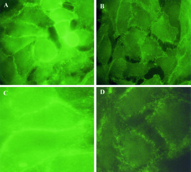
In the present study, we evaluated whether motility of Kaposi's sarcoma (KS) cells induced by platelet-activating factor (PAF) is dependent on the regulation of adherens junctions components. The results obtained indicate that PAF dose and time dependently reduced the endogenous expression of the main components of the adherens junctions: VE-cadherin, alpha-catenin, and beta-catenin. In addition, PAF initiated events that directly or indirectly up-regulated both the tyrosine and serine/threonine phosphorylation pathways, and both types of phosphorylation of beta-catenin were involved in the motility of KS cells. This motility was abrogated by addition of the tyrosine kinase inhibitor genistein, suggesting that this phosphorylation is an important signal responsible for breaking down the adherens junctions and diminishing the ability of neighboring cells to interact. Furthermore, immunofluorescence analysis showed that beta-catenin and VE-cadherin staining changed from a uniform distribution along the membrane of controls to a diffuse pattern with gap formation in PAF-treated KS cells. In conclusion, the data presented here indicate that PAF induces tumor cell motility by altering cell-cell adhesion through beta-catenin phosphorylation.


Figures

Figure 1
Figure 1
The regulation of total protein expression of VE-cadherin in KS cells by PAF. A: Time course of 50 ng/ml of PAF on VE-cadherin expression. Cells were incubated with vehicle alone or with PAF at the indicated concentration for varying time points up to 12 hours; this is summarized by densitometric analysis (bottom) showing the decrease of its expression in total cell lysate. B: Dose response of PAF on VE-cadherin expression; cells were stimulated for 4 hours with or without various concentration of PAF (1 to 50 ng/ml), which is summarized graphically (bottom). C, cells incubated with vehicle alone. The data shown are representative of three different experiments.
Figure 2
Figure 2
The inhibitory effect of specific PAF receptor antagonists Ginkgolide B or CV 3988 on VE-cadherin expression. Cells were incubated with PAF (10 ng/ml) in the presence or absence of 0.34 μmol/L Ginkgolide B (G) or 3 μmol/L CV 3988 (CV) for 4 hours. C, cells incubated with the vehicle alone. The data shown are representative of three different experiments.
Figure 3
Figure 3
The regulation of total protein expression of β-catenin in KS cells by PAF. Cells were stimulated with the vehicle alone (C) or with PAF (10 ng/ml) or PAF plus PAF receptor antagonists Ginkgolide B (G; 0.34 μmol/L) or CV 3988 (CV; 3 μmol/L) for time ranging from 30 minutes to 12 hours. Top: The blot after immunoprobing with a polyclonal β-catenin antibody. Bottom: The quantitative measurements of the band intensities. The data shown are representative of three different experiments.
Figure 4
Figure 4
PAF regulates total protein expression of α-catenin in KS cells as assessed by Western blotting. Cells were stimulated with vehicle alone (C) or with PAF (10 ng/ml) or PAF plus PAF receptor antagonists Ginkgolide B (G; 0.34 μmol/L) or CV 3988 (CV; 3 μmol/L) at the indicated times. Top: The blot after immunoprobing with a polyclonal α-catenin antibody, which is summarized in the bottom quantitative graph. The data shown are representative of three different experiments.
Figure 5
Figure 5
PAF stimulates phosphorylation of β-catenin in KS cells. Subconfluent cultures of KS cells were starved for 12 hours and then incubated with PAF (10 and 50 ng/ml) at the indicated times. Cells were also treated with the vehicle alone (C), or with PAF (50 ng/ml) plus PAF-receptor antagonists Ginkgolide B (G; 0.34 μmol/L) or CV 3988 (CV; 3 μmol/L) for 1 hour. A: Cells extracts were subject to immunoprecipitation with antibody against β-catenin. Precipitated proteins were analyzed by SDS-PAGE followed by immunoblotting with an antibody to phosphotyrosine. PY-β-catenin, tyrosine phosphorylated β-catenin. B: Western blotting analysis using the anti-phospho-S33/S37/T41 antibody to β-catenin. The data shown are representative of three different experiments.
Figure 6
Figure 6
Effects of PAF on ubiquitination of β-catenin. KS cells were cultured for the indicated times in the absence (C) or presence of 10 ng/ml of PAF. Then cellular proteins were extracted and immunoprecipitated with an anti-β-catenin antibody as described in the Materials and Methods section. The immunoprecipitate was subsequently run on SDS/PAGE and immunoblotted for ubiquitin. The experiment is representative of at least three different experiments giving similar results. Ub β-cat, ubiquitinated β-catenin.
Figure 7
Figure 7
Effect of PAF on the junctional localization of β-catenin and VE-cadherin in KS cells. Cells were grown to subconfluence and treated with 10 ng/ml of PAF for 2 hours. A and C represent control staining for β-catenin and VE-cadherin, respectively; B and D represent β-catenin and VE-cadherin staining after PAF treatment. In B and D, it is evident the gap formation and absence of β-catenin and VE-cadherin in areas where the cells have separated. PAF also promoted a zigzag pattern distribution of β-catenin (B) and a diffuse staining pattern for VE-cadherin (D) different from controls displaying a linear pattern distribution localized at cell-cell contact. Three experiments were performed with similar results. Original magnifications, ×400.
Figure 8
Figure 8
Effect of tyrosine kinase inhibitor genistein on PAF-induced KS motility. Cells were plated on 35 × 10-mm tissue culture dishes in RPMI medium with 0.25% BSA and then were incubated with vehicle alone (C) or with 10 ng/ml of PAF or with PAF plus genistein (100 μmol/L) or with PAF plus the PAF-receptor antagonist CV 3988 (CV; 3 μmol/L). For inhibition studies, genistein was added 1 hour before the addition of PAF. Cell motility was monitored with a time-lapse cinematography system as described in the Materials and Methods section. The values are the average of triplicate cultures ± SD. Inset: Cell treated with PAF plus genistein (100 μmol/L) and stained for β-catenin.

Similar articles

Cited by

References

    1. Prescott SM, Zimmerman GA, McIntyre TM. Platelet-activating factor. J Biol Chem. 1990;265:17381–17384. - PubMed
    1. Kume K, Shimizu T. Platelet-activating factor (PAF) induces growth stimulation, inhibition, and suppression of oncogenic transformation in NRK cells overexpressing the PAF receptor. J Biol Chem. 1997;272:22898–22904. - PubMed
    1. Boccellino M, Biancone L, Cantaluppi V, Ye RD, Camussi G. Effect of platelet-activating factor receptor expression on CHO cell motility. J Cell Physiol. 2000;183:254–264. - PubMed
    1. Biancone L, Cantaluppi V, Boccellino M, Bussolati B, Del Sorbo L, Conaldi PG, Albini A, Toniolo A, Camussi G. Motility induced by human immunodeficiency virus-1 Tat on Kaposi’s sarcoma cells requires platelet-activating factor synthesis. Am J Pathol. 1999;155:1731–1739. - PMC - PubMed
    1. Takeichi M. Cadherin cell adhesion receptors as a morphogenetic regulator. Science. 1991;251:1451–1455. - PubMed

Publication types