

Expression profiling of primary tumors and matched lymphatic and lung metastases in a xenogeneic breast cancer model
- PMID: 15855655
- PMCID: PMC1606408
- DOI: 10.1016/S0002-9440(10)62372-3
Expression profiling of primary tumors and matched lymphatic and lung metastases in a xenogeneic breast cancer model
Abstract
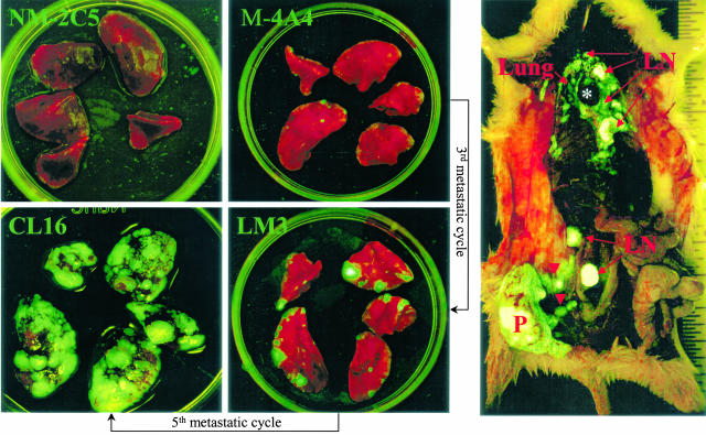
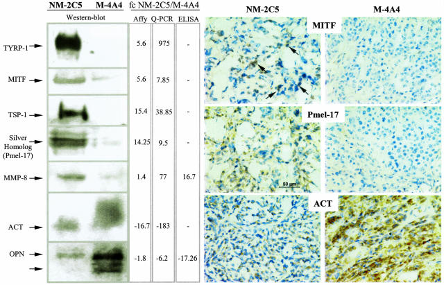
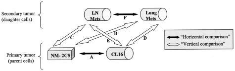
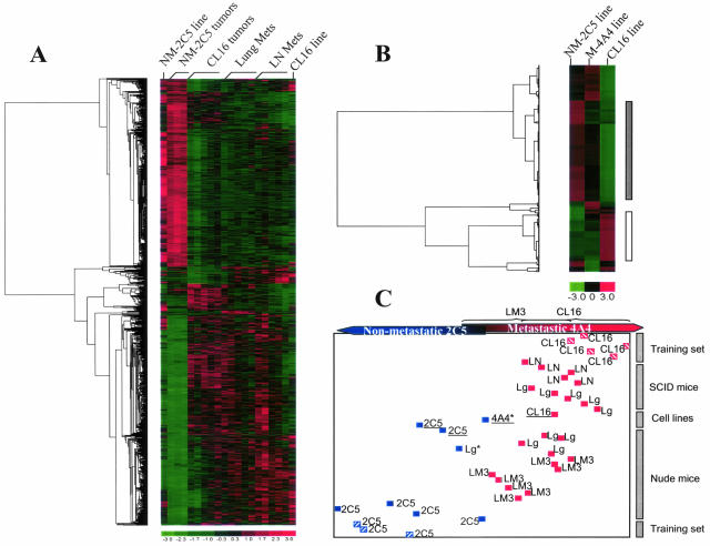
Using a purpose-designed experimental model, we have defined new, statistically significant, differences in gene expression between heavily and weakly metastatic human breast cancer cell populations, in vivo and in vitro. The differences increased under selection pressures designed to increase metastatic proficiency. Conversely, the expression signatures of primary tumors generated by more aggressive variants, and their matched metastases in the lungs and lymph nodes, all tended to converge. However, the few persisting differences among these selectively enriched malignant growths in the breast, lungs, and lymph nodes were highly statistically significant, implying potential mechanistic involvement of the corresponding genes. The evidence that has emerged from the current work indicates that selective enhancement of metastatic proficiency by serial transplantation co-purifies a subliminal gene expression pattern within the tumor cell population. This signature most likely includes genes participating in metastasis pathogenesis, and we document manageable numbers of candidates for this role. The findings also suggest that metastasis to at least two different organs occurs through closely similar genetic mechanisms.
Figures
Comment in
-
New insights into the tumor metastatic process revealed by gene expression profiling.Am J Pathol. 2005 May;166(5):1291-4. doi: 10.1016/S0002-9440(10)62348-6. Am J Pathol. 2005. PMID: 15855631 Free PMC article. Review. No abstract available.
Similar articles
-
Analysis of paired primary lung and lymph node tumor cells: a model of metastatic potential by multiple genetic programs.Cancer Detect Prev. 2005;29(6):509-17. doi: 10.1016/j.cdp.2005.09.006. Epub 2005 Nov 10. Cancer Detect Prev. 2005. PMID: 16289389
-
Bcl-x(L)-mediated changes in metabolic pathways of breast cancer cells: from survival in the blood stream to organ-specific metastasis.Am J Pathol. 2005 Oct;167(4):1125-37. doi: 10.1016/S0002-9440(10)61201-1. Am J Pathol. 2005. PMID: 16192647 Free PMC article.
-
Using a xenograft model of human breast cancer metastasis to find genes associated with clinically aggressive disease.Cancer Res. 2005 Jul 1;65(13):5578-87. doi: 10.1158/0008-5472.CAN-05-0108. Cancer Res. 2005. PMID: 15994930
-
A six-gene signature predicting breast cancer lung metastasis.Cancer Res. 2008 Aug 1;68(15):6092-9. doi: 10.1158/0008-5472.CAN-08-0436. Cancer Res. 2008. PMID: 18676831
-
[Identification of the differentially expressed genes between primary breast cancer and paired lymph node metastasis through combining mRNA differential display and gene microarray].Zhonghua Yi Xue Za Zhi. 2006 Oct 24;86(39):2749-55. Zhonghua Yi Xue Za Zhi. 2006. PMID: 17199993 Chinese.
Cited by
-
Biomarkers of residual disease, disseminated tumor cells, and metastases in the MMTV-PyMT breast cancer model.PLoS One. 2013;8(3):e58183. doi: 10.1371/journal.pone.0058183. Epub 2013 Mar 8. PLoS One. 2013. PMID: 23520493 Free PMC article.
-
High Rab27A expression indicates favorable prognosis in CRC.Diagn Pathol. 2015 Jun 13;10:68. doi: 10.1186/s13000-015-0303-3. Diagn Pathol. 2015. PMID: 26070933 Free PMC article.
-
Cancer invasion and metastasis: interacting ecosystems.Virchows Arch. 2009 Jun;454(6):599-622. doi: 10.1007/s00428-009-0784-0. Epub 2009 May 27. Virchows Arch. 2009. PMID: 19471961 Review.
-
The molecular profile of breast cancer: primary tumor versus corresponding lymph node metastases.Rom J Morphol Embryol. 2022 Apr-Jun;63(2):421-429. doi: 10.47162/RJME.63.2.13. Rom J Morphol Embryol. 2022. PMID: 36374147 Free PMC article.
-
NSG Mice Provide a Better Spontaneous Model of Breast Cancer Metastasis than Athymic (Nude) Mice.PLoS One. 2016 Sep 23;11(9):e0163521. doi: 10.1371/journal.pone.0163521. eCollection 2016. PLoS One. 2016. PMID: 27662655 Free PMC article.
References
-
- Fidler IJ, Kripke ML. Metastasis results from preexisting variant cells within a malignant tumor. Science. 1977;197:893–895. - PubMed
-
- Fidler IJ. Tumor heterogeneity and the biology of cancer invasion and metastasis. Cancer Res. 1978;38:2651–2660. - PubMed
-
- Fidler IJ. The pathogenesis of cancer metastasis: the ‘seed and soil’ hypothesis revisited. Nat Rev Cancer. 2003;3:453–458. - PubMed
-
- Price JE, Carr D, Tarin D. Spontaneous and induced metastasis of naturally occurring tumors in mice: analysis of cell shedding into the blood. J Natl Cancer Inst. 1984;73:1319–1326. - PubMed
-
- Tarin D, Price JE, Kettlewell MG, Souter RG, Vass AC, Crossley B. Mechanisms of human tumor metastasis studied in patients with peritoneovenous shunts. Cancer Res. 1984;44:3584–3592. - PubMed
Publication types
MeSH terms
LinkOut - more resources
Full Text Sources
Other Literature Sources
Medical
