. 2005 Jul 28;24(32):5053-68.
doi: 10.1038/sj.onc.1208685.

Loss of TGF-beta type II receptor in fibroblasts promotes mammary carcinoma growth and invasion through upregulation of TGF-alpha-, MSP- and HGF-mediated signaling networks

Affiliations

Loss of TGF-beta type II receptor in fibroblasts promotes mammary carcinoma growth and invasion through upregulation of TGF-alpha-, MSP- and HGF-mediated signaling networks

Nikki Cheng et al. Oncogene. .

Abstract

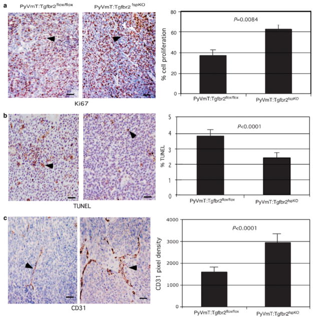
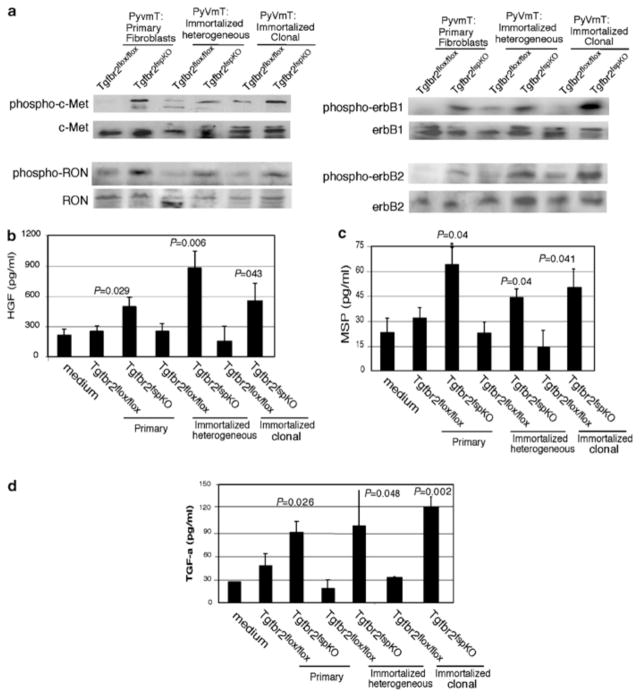
Stromal fibroblasts regulate epithelial cell behavior through direct and indirect cell-cell interactions. To clarify the role of TGF-beta signaling in stromal fibroblasts during mammary development and tumorigenesis, we conditionally knocked out the TGF-beta type II receptor gene in mouse mammary fibroblasts (Tgfbr2(fspKO)). Tgfbr2(fspKO) mice exhibit defective mammary ductal development, characterized in part by increased ductal epithelial cell turnover associated with an increase in stromal fibroblast abundance. Tgfbr2(fspKO) mammary fibroblasts transplanted with mammary carcinoma cells promote growth and invasion, which is associated with increased activating phosphorylation of the receptors: erbB1, erbB2, RON, and c-Met. Furthermore, the increased receptor phosphorylation correlates with increased secretion of the cognate ligands by Tgfbr2(fspKO) fibroblasts. Treatment of tumor cells with fibroblast-conditioned medium leads to increased tumor cell proliferation and motility, which are blocked by addition of pharmacologic inhibitors of TGF-alpha signaling or neutralizing antibodies to macrophage-stimulating protein (MSP), HGF, or c-Met. These studies characterize a significant role for stromal TGF-beta signaling in mammary tissue homeostasis and mammary tumor progression via regulation of TGF-alpha, MSP, and HGF signaling pathways.


Figures

Figure 1
Figure 1
FSP1 promoter activity is exclusive to fibroblasts in the mammary gland. (a) Analysis of GFP expression in the stromal cells of mammary tissue from FSP.GFP transgenic reporter mice as visualized under fluorescence microscopy. GFP-expressing cells are indicated by arrowheads. Non-GFP-expressing stromal cells are indicated by arrows. (b) Histological analysis of lacZ staining in stromal cells from mammary tissue sections of ROSA26A Cre reporter mice as indicated by arrowheads. Non-lacZ-stained stromal cells are indicated by arrows. Scale bars represent 40 μm. (c) FSP.Cre-mediated recombination in fibroblasts was determined by PCR analyses of DNA extracted from the mammary stromal tissue of Tgfbr2fspKO and control mice. Stromal tissue was micro-dissected by laser capture from mammary tissue sections as indicated by arrowheads. Left and right panels indicate before and after laser capture microdissection, respectively. DNA, extracted for PCR analysis, was used to address recombination of the Tgfbr2 allele in the mammary stroma of Tgfbr2fspKO mice, as indicated by arrowheads. The positive control was generated using DNA extracted from PyVmT cells containing floxed Tgfbr2 and expressing Cre under the control of the MMTV promoter. (d) Recombination of Tgfbr2 alleles in cultured mammary fibroblasts was verified by PCR. (e) Specificity of FSP.Cre-mediated recombination was determined by PCR analyses of DNA extracted from mammary stromal (S) and epithelial (E) tissue of Tgfbr2fspKO mice. Epithelial tissue was micro-dissected by laser capture from mammary tissue sections as indicated by arrowheads and subjected to PCR analyses for recombination of the Tgfbr2 allele. Left and right panels indicate before and after laser capture microdissection, respectively. The positive control was generated using DNA extracted from PyVmT cells containing floxed Tgfbr2 and expressing Cre under the control of the MMTV promoter. (f) Specificity of recombination of Tgfbr2 alleles was confirmed by PCR analyses on DNA extracted from cultured mammary fibroblasts and epithelial cells. The positive control was generated using DNA extracted from PyVmT cells containing floxed Tgfbr2 and expressing Cre under the control of the MMTV promoter. The negative control was generated using DNA extracted from PyVmT cells containing floxed Tgfbr2 and not expressing Cre.
Figure 2
Figure 2
Tgfbr2fspKO mice exhibit severe defects in mammary gland development. (a) Whole mounts of mammary gland tissue isolated from female Tgfbr2fspKO and Tgfbr2flox/flox mice at 6 weeks of age, showing differences in ductal development at low and high magnifications. In the Tgfbr2flox/flox, ducts have extended well beyond the central lymph node (*), but this is not the case in Tgfbr2fspKO mammary glands. Arrowheads indicate terminal end buds. (b) A significant increase in the number of fibroblasts was detected in the mammary glands of Tgfbr2fspKO.GFP mice (n=15), compared to Tgfbr2flox/flox mice (n=15), which was determined by quantification of FSP.GFP-expressing cells. Red oval indicates mean. (c) Quantitation of Ki67 immunostaining of mammary gland tissue from mice at 6 weeks of age indicates an increase in cell proliferation in the ductal epithelium (n=20) of Tgfbr2fspKO mice compared to Tgfbr2flox/flox (n=20). A decrease in cell proliferation was detected in the terminal end bud cells of Tgfbr2fspKO mice (n=13) compared to Tgfbr2flox/flox mice (n=16). Red oval indicates the mean. Statistical significance was determined two-tailed Student’s t-test. (d) Quantitation of cleaved caspase 3 immunostaining of mammary ducts and terminal ends of mammary gland tissue isolated from mice at 6 weeks of age indicate increase in ductal epithelial apoptosis in Tgfbr2fspKO mice (n=8) compared to Tgfbr2flox/flox mice (n=6). No significant change in apoptosis was detected in terminal end bud cells in Tgfbr2fspKO mice (n=16) compared to Tgfbr2flox/flox mice (n=16). Red oval indicates the mean. Statistical significance was determined by two-tailed Student’s t-test.
Figure 3
Figure 3
Tgfbr2fspKO fibroblasts exhibit defective responses to TGF-β signaling. (a) Southern blot analysis of mammary fibroblast DNA showing recombination efficiencies of Tgfbr2 floxed alleles of DNA samples from primary fibroblasts (lanes 1–3), immortalized heterogenous (lanes 4–5), and immortalized clonal fibroblasts (lanes 6–7). One cell line representative of three Tgfbr2flox/flox and of three Tgfbr2fspKO clones examined is shown here. Numbered lanes are as follows: (1) Tgfbr2flox/flox, (2) Tgfbr2fspKO, (3) TgfbrfspKO, (4) Tgfbr2flox/flox, (5) Tgfbr2fspKO, (6) Tgfbr2flox/flox, (7) Tgfbr2fspKO, (8) control for Tgfbr2flox/flox, (9) control for Tgfbr2fspKO. Control DNA samples were isolated from MMTV.PyVmT transgenic mice expressing MMTV-Cre with Floxed Tgfbr2. Recombination efficiency was determined by phosphorimager analyses. (b) Primary or immortalized mammary fibroblasts from Tgfbr2flox/flox and Tgfbr2fspKO mice were treated with TGF-β and subjected to 3[H]thymidine incorporation. One representative of three Tgfbr2flox/flox and three Tgfbr2fspKO clones is shown here. (c) Primary or (d, e) immortalized fibroblasts from Tgfbr2flox/flox and Tgfbr2fspKO mice were treated with TGF-β, and counted by a hemocytometer at the indicated time-points.
Figure 4
Figure 4
Subrenal capsule grafting of PyVmT tumor cells with Tgfbr2fspKO fibroblasts result in increased tumor growth and invasion in vivo. (a) H&E staining of tumor tissue isolated 30 days after grafting showing tumor invasion into the kidney parenchyma. Scale bars represent 40 μm. (b) Exogenous fibroblasts, not endogenous fibroblasts, are primary contributors to tumor growth. Tgfbr2flox/flox and Tgfbr2fspKO cells were labeled with CMFDA flourochrome and grafted with PyVmT. Tumors were harvested 30 days after grafting, formalin fixed and stained by H&E. Fibroblasts were visualized by fluorescent microscopy. To control for background fluorescence, sections, sections containing labeled fibroblasts were compared to tumor sections containing unlabeled fibroblasts; the latter showed no fluorescence. Scale bars represent 40 μm.
Figure 5
Figure 5
PyVmT grafted with Tgfbr2fspKO cells exhibit increased tumor angiogenesis, increased proliferation and decreased apoptosis. Tumor tissue was harvested 30 days post grafting, formalin fixed and subjected in immunohistochemical staining for (a) Ki67 (b) apoptotic nuclei by TUNEL and (c). CD31 expression as indicated by arrowheads. Quantitation of CD31 immunostaining was determined by measuring pixel density using Scion Image Software of five fields of two sections per sample (n=18). Quantitation of Ki67 and TUNEL immunostaining were determined by counting number of peroxidase stained nuclei over the total number of hematoxylin-stained nuclei in five fields of two sections per sample (n=18). Scale bars represent 40 μm. Quantitation of PyVmT cells grafted with immortalized heterogeneous fibroblasts is depicted here. Statistical significance was determined by Poisson regression.
Figure 6
Figure 6
Tgfbr2fspKO fibroblasts increased receptor tyrosine kinase phosphorylation in PyVmT tumors. Cell lysates prepared from 30-day grafts containing PyVmT cells and fibroblasts were subjected to Western blot analysis for: (a) phosphorylation of the indicated cell surface receptors; (b) levels of soluble TGF-α, (c) MSP, and (d). HGF in conditioned media from primary and immortalized Tgfbr2flox/flox and Tgfbr2fspKO fibroblasts were determined by ELISA. Statistical significance was determined by two-tailed Student’s t-test.
Figure 7
Figure 7
Elevated levels of TGF-α, MSP, and HGF secretion by Tgfbr2fspKO fibroblasts contribute to PyVmT proliferation and migration. (a) PyVmT cells were treated with fibroblast-conditioned medium from Tgfbr2flox/flox and Tgfbr2fspKO clonal fibroblasts in the presence or absence of gefitinib, or neutralizing antibodies to HGF, c-Met, or MSP for 24 h, and proliferation was measured by [3H]thymidine incorporation. Statistical comparison of proliferation between treatment of PyVmT tumor cells with conditioned medium from Tgfbr2flox/flox and from Tgfbr2fspKO clonal fibroblasts was determined by two-tailed Student’s t-test. P-value significance is indicated by *. (b) PyVmT cells were wounded, and then treated with fibroblast-conditioned medium from Tgfbr2flox/flox and Tgfbr2fspKO clonal fibroblasts in the presence or absence of gefitinib, or neutralizing antibodies to HGF, c-Met, or MSP. Migration was assessed at 12 h by wound closure using Scion Image software. Results of conditioned medium experiments from one Tgfbr2flox/flox and one Tgfbr2fspKO cell line are depicted here and are representative of three Tgfbr2flox/flox and three Tgfbr2fspKO cell lines examined. Statistical comparison of migration between treatment of PyVmT with conditioned medium from Tgfbr2flox/flox and Tgfbr2fspKO clonal fibroblasts was determined by two-tailed Student’s t-test. P-value significance is indicated by *.

References

    1. Akhurst RJ. J Clin Invest. 2002;109:1533–1536. - PMC - PubMed
    1. Akhurst RJ, Derynck R. Trends Cell Biol. 2001;11:S44–S50. - PubMed
    1. Aslakson C, Miller FR. Cancer Res. 1992;52:1399–1405. - PubMed
    1. Bhowmick N, Chytiil A, Plieth D, Gorska A, Dumont N, Shappel S, Washington M, Neilson E, Moses H. Science. 2004;303:847–851. - PubMed
    1. Bitzer M, von Gersdorff G, Liang D, Dominguez-Rosales A, Beg AA, Rojkind M, Bottinger EP. Genes Dev. 2000;2:187–197. - PMC - PubMed

Publication types

MeSH terms