

Genome-wide gene expression profiling of testicular carcinoma in situ progression into overt tumours
- PMID: 15856041
- PMCID: PMC2361756
- DOI: 10.1038/sj.bjc.6602560
Genome-wide gene expression profiling of testicular carcinoma in situ progression into overt tumours
Abstract
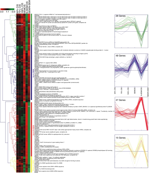
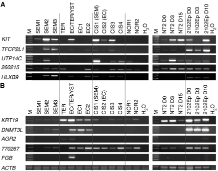
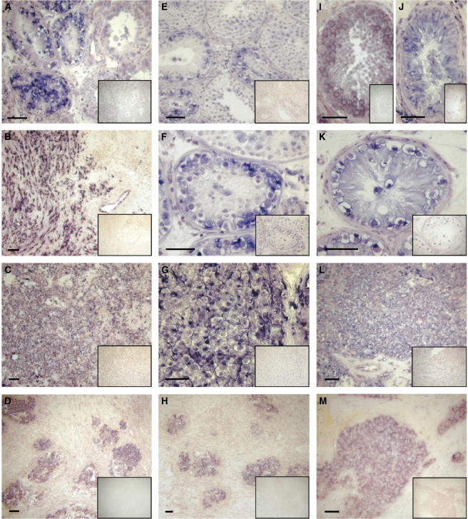
The carcinoma in situ (CIS) cell is the common precursor of nearly all testicular germ cell tumours (TGCT). In a previous study, we examined the gene expression profile of CIS cells and found many features common to embryonic stem cells indicating that initiation of neoplastic transformation into CIS occurs early during foetal life. Progression into an overt tumour, however, typically first happens after puberty, where CIS cells transform into either a seminoma (SEM) or a nonseminoma (N-SEM). Here, we have compared the genome-wide gene expression of CIS cells to that of testicular SEM and a sample containing a mixture of N-SEM components, and analyse the data together with the previously published data on CIS. Genes showing expression in the SEM or N-SEM were selected, in order to identify gene expression markers associated with the progression of CIS cells. The identified markers were verified by reverse transcriptase-polymerase chain reaction and in situ hybridisation in a range of different TGCT samples. Verification showed some interpatient variation, but combined analysis of a range of the identified markers may discriminate TGCT samples as SEMs or N-SEMs. Of particular interest, we found that both DNMT3B (DNA (cytosine-5-)-methyltransferase 3 beta) and DNMT3L (DNA (cytosine-5-)-methyltransferase 3 like) were overexpressed in the N-SEMs, indicating the epigenetic differences between N-SEMs and classical SEM.
Figures
References
-
- Adami HO, Bergstrom R, Mohner M, Zatonski W, Storm H, Ekbom A, Tretli S, Teppo L, Ziegler H, Rahu M, Gurevicius R, Stengrevics A (1994) Testicular cancer in nine northern European countries. Int J Cancer 59: 33–38 - PubMed
-
- Albrechtsen R, Nielsen MH, Skakkebaek NE, Wewer U (1982) Carcinoma in situ of the testis. Some ultrastructural characteristics of germ cells. Acta Pathol Microbiol Immunol Scand [A] 90: 301–303 - PubMed
-
- Almstrup K, Hoei-Hansen CE, Wirkner U, Blake J, Schwager C, Ansorge W, Nielsen JE, Skakkebaek NE, Rajpert-De Meyts E, Leffers H (2004) Embryonic stem cell-like features of testicular carcinoma in situ revealed by genome-wide gene expression profiling. Cancer Res 64: 4736–4743 - PubMed
-
- Andrews PW (1984) Retinoic acid induces neuronal differentiation of a cloned human embryonal carcinoma cell line in vitro. Dev Biol 103: 285–293 - PubMed
-
- Andrews PW, Banting G, Damjanov I, Arnaud D, Avner P (1984) Three monoclonal antibodies defining distinct differentiation antigens associated with different high molecular weight polypeptides on the surface of human embryonal carcinoma cells. Hybridoma 3: 347–361 - PubMed
Publication types
MeSH terms
Substances
LinkOut - more resources
Full Text Sources
Medical
