

Reduced coronary flow reserve in Anderson-Fabry disease measured by transthoracic Doppler echocardiography
- PMID: 15857518
- PMCID: PMC1097744
- DOI: 10.1186/1476-7120-3-11
Reduced coronary flow reserve in Anderson-Fabry disease measured by transthoracic Doppler echocardiography
Abstract
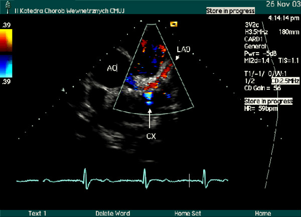
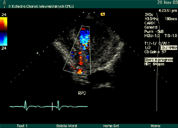
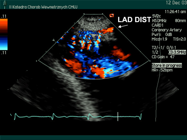
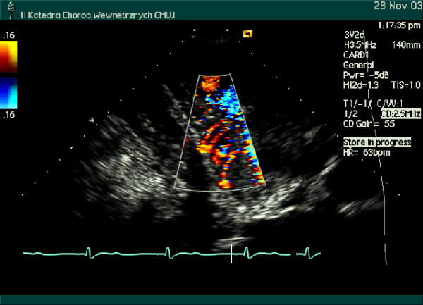
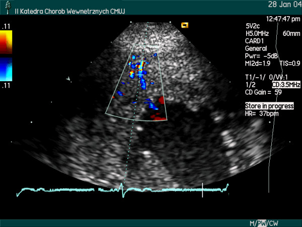
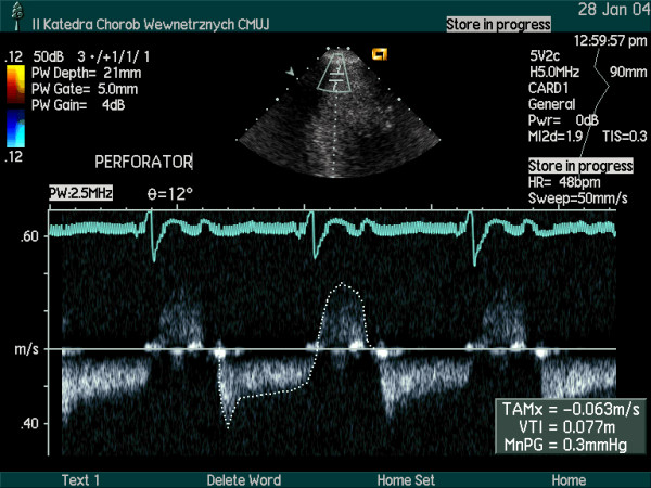
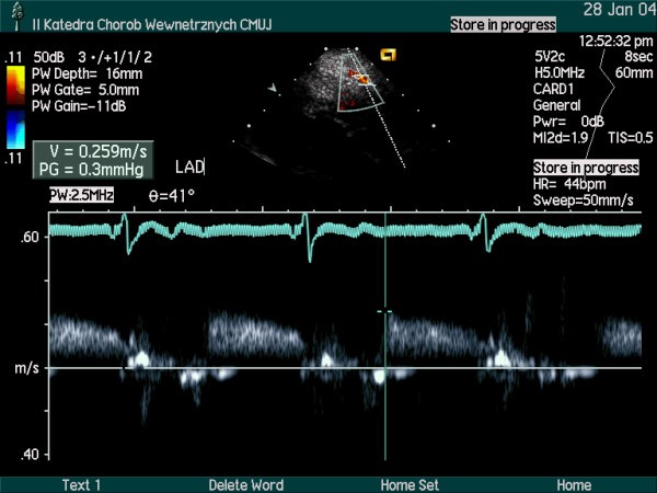
Coronary flow reserve was assessed in a patient with Anderson-Fabry disease complicated by symmetric left ventricular hypertrophy. Coronary flow reserve was measurable in all three major coronary arteries providing an opportunity to compare regional coronary flow reserve from different vascular beds. In this patient all the three vascular beds supplied diffusely hypertrophied myocardium. Coronary flow disturbances in small intramyocardial perforating arteries were visible. The coronary flow reserve was reduced to a similar level (around to 2.0) in all three major arteries. In our patient with Anderson-Fabry disease, the coronary vasodilatation was blunted in a diffuse pattern corresponding to the myocardial hypertrophy distribution. In small intramyocardial arteries coronary flow was also disturbed. Accordingly, retrograde systolic flow and accelerated anterograde diastolic flow were documented.
Figures
Similar articles
-
Demonstration of penetrating intramyocardial coronary arteries with high-frequency transthoracic echocardiography and Doppler in human subjects.J Am Soc Echocardiogr. 1999 Jan;12(1):55-63. doi: 10.1016/s0894-7317(99)70173-2. J Am Soc Echocardiogr. 1999. PMID: 9882779
-
Transthoracic Doppler echocardiographic assessment of left anterior descending coronary artery and intramyocardial small coronary artery flow in patients with hypertrophic cardiomyopathy.J Cardiol. 2001;37 Suppl 1:115-20. J Cardiol. 2001. PMID: 11433814
-
New noninvasive diagnosis of myocardial ischemia of the left circumflex coronary artery using coronary flow reserve measurement by transthoracic Doppler echocardiography: comparison with thallium-201 single photon emission computed tomography.J Cardiol. 2004 Mar;43(3):109-16. J Cardiol. 2004. PMID: 15067798
-
Transthoracic Doppler echocardiography - noninvasive diagnostic window for coronary flow reserve assessment.Cardiovasc Ultrasound. 2003 Apr 11;1:4. doi: 10.1186/1476-7120-1-4. Cardiovasc Ultrasound. 2003. PMID: 12740038 Free PMC article. Review.
-
Demonstration of coronary artery flow using transthoracic Doppler echocardiography.J Am Soc Echocardiogr. 2004 Feb;17(2):178-85. doi: 10.1016/j.echo.2003.08.017. J Am Soc Echocardiogr. 2004. PMID: 14752494 Review.
Cited by
-
Effect of enzyme replacement therapy on serum asymmetric dimethylarginine levels, coronary flow reserve and left ventricular hypertrophy in patients with Fabry disease.Clin Kidney J. 2012 Dec;5(6):512-518. doi: 10.1093/ckj/sfs114. Epub 2012 Oct 3. Clin Kidney J. 2012. PMID: 24976967 Free PMC article.
-
Establishing 3-nitrotyrosine as a biomarker for the vasculopathy of Fabry disease.Kidney Int. 2014 Jul;86(1):58-66. doi: 10.1038/ki.2013.520. Epub 2014 Jan 8. Kidney Int. 2014. PMID: 24402087 Free PMC article.
-
Inflammation, Oxidative Stress, and Endothelial Dysfunction in the Pathogenesis of Vascular Damage: Unraveling Novel Cardiovascular Risk Factors in Fabry Disease.Int J Mol Sci. 2024 Jul 29;25(15):8273. doi: 10.3390/ijms25158273. Int J Mol Sci. 2024. PMID: 39125842 Free PMC article. Review.
-
Status and potential clinical value of a transthoracic evaluation of the coronary arteries.Cardiovasc Ultrasound. 2016 Jan 19;14:5. doi: 10.1186/s12947-016-0048-5. Cardiovasc Ultrasound. 2016. PMID: 26787070 Free PMC article. Review.
-
Biomarkers in Fabry Disease. Implications for Clinical Diagnosis and Follow-up.J Clin Med. 2021 Apr 13;10(8):1664. doi: 10.3390/jcm10081664. J Clin Med. 2021. PMID: 33924567 Free PMC article. Review.
References
Publication types
MeSH terms
LinkOut - more resources
Full Text Sources
Medical
