. 2005;132(4):943-53.
doi: 10.1016/j.neuroscience.2005.01.020.

Infralimbic cortex activation increases c-Fos expression in intercalated neurons of the amygdala

Affiliations

Infralimbic cortex activation increases c-Fos expression in intercalated neurons of the amygdala

S Berretta et al. Neuroscience. 2005.

Abstract

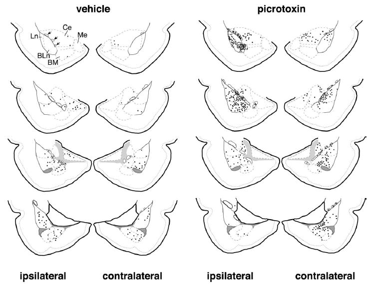
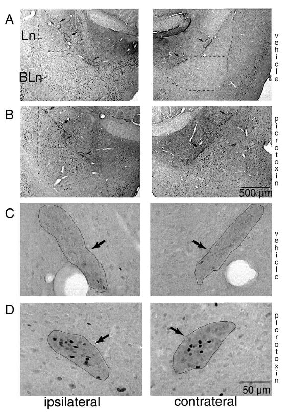
Recently, it was reported that stimulation of the infralimbic cortex produces a feedforward inhibition of central amygdala neurons. The interest of this observation comes from the fact that the central nucleus is the main output station of the amygdala for conditioned fear responses and evidence that the infralimbic cortex plays a critical role in the extinction of conditioned fear. However, the identity of the neurons mediating this infralimbic-evoked inhibition of the central nucleus remains unknown. Likely candidates are intercalated amygdala neurons. Indeed, these cells receive glutamatergic afferents from the infralimbic cortex, use GABA as a transmitter, and project to the central amygdala. Thus, the present study was undertaken to test whether, in adult rats, the infralimbic cortex can affect the activity of intercalated neurons. To this end, disinhibition of the infralimbic cortex was induced by local infusion of the non-competitive GABA-A receptor antagonist picrotoxin. Subsequently, neuronal activation was determined bilaterally within the amygdala using induction of the immediate early gene Fos. Infralimbic disinhibition produced a significant increase in the number of Fos-immunoreactive intercalated cells bilaterally whereas no change was detected in the central nucleus. In the basolateral amygdaloid complex, increases in the number of Fos-immunoreactive cells only reached significance in the contralateral lateral nucleus. These results suggest that glutamatergic inputs from the infralimbic cortex directly activate intercalated neurons. Thus, our findings raise the possibility that the infralimbic cortex inhibits conditioned fear via the excitation of intercalated cells and the consequent inhibition of central amygdala neurons.


Figures

Fig. 1
Fig. 1
Histological determination of infusion sites in the prefrontal cortex. Injection cannulas were aimed at the superficial layers of the infralimbic cortex. In A, a microphotograph of a Cresyl Violet-stained brain section from a rat infused with picrotoxin. The arrow indicates the tip of the cannula track, located within the superficial layers of the infralimbic cortex. In B, diagrammatic representations of two levels of the prefrontal cortex (modified from Swanson, 1992) indicating the tip of the cannula tracks for saline- (triangles) and picrotoxin- (stars) injected rats. Solid symbols mark successful infralimbic injections. Dotted symbols represent five cases that were excluded from the final analysis because the tip of the cannula track lay outside the infralimbic cortex. Abbreviations: IL, infralimbic cortex; PL, prelimbic cortex.
Fig. 2
Fig. 2
Distribution of Fos-IR cells in rats that received vehicle vs. picrotoxin injections in the infralimbic cortex. Picrotoxin infusion in the infralimbic cortex was associated with an increase of Fos induction overall in the amygdala. Such increase was particularly noticeable within the ITC cell masses, both ipsi- and contra-laterally respect to the injection site. Diagrammatic representations of the amygdala (modified from Swanson, 1992) are shown for representative vehicle- (left) and picrotoxin- (right) treated animals. Sections are displayed in rostro-caudal order from top to bottom. Each black dot represents three Fos-IR nuclei. ITC cell masses are outlined with a continuous line and are marked by arrows in the upper left diagram, while the borders of the other amygdala nuclei are marked with a dashed line. BLn, basolateral nucleus; BM, basomedial nucleus; Ce, central nucleus; Ln, lateral nucleus; Me, medial nucleus.
Fig. 3
Fig. 3
GABA and Fos immunoreactivity in the ITC cell masses. Low (A, B) and high (C, D) magnification microphotographs of brain sections processed for dual antigen immunocytochemistry for GABA and Fos. The borders of the Ln and BLn are indicated with a dashed line while the ITC cell masses are outlined with a continuous line and marked with arrows. Fos-IR nuclei can be seen as darkly stained cell nuclei. GABA immunostaining was detectable as a diffuse neuropil labeling that was sufficiently intense to allow a reliable delineation of the ITC cell masses even though individual cell bodies were only sporadically detected. Sections shown in A (vehicle) and B (picrotoxin) are typical for distribution of Fos-IR nuclei within the amygdalar subnuclei. Statistically significant increases were only detected in the ITC cells masses and in the contralateral lateral nucleus of picrotoxin-treated rats. In C (vehicle) and D (picrotoxin), examples of ITC cell masses are outlined in black. Note the numerous Fos-IR nuclei in the rat treated with picrotoxin (D). BLn, basolateral nucleus; BM, basomedial nucleus; Ce, central nucleus; Ln, lateral nucleus; Me, medial nucleus.
Fig. 4
Fig. 4
Effect of infralimbic picrotoxin infusions on the number Fos-IR cells in the ITC cell masses. Significant increases in the total number of Fos-IR nuclei were detected in the rostral (ipsilaterally) and caudal (contralaterally) ITC cell masses in rats infused with picrotoxin within the infralimbic cortex.
Fig. 5
Fig. 5
Effect of infralimbic picrotoxin infusions on the densities of Fos-IR cells in the ipsilateral Ln, BLnM, BLnP, CeL and CeM of the amygdala. No significant changes of densities of Fos-IR nuclei were detected ipsilaterally in any of the regions examined. In both the picrotoxin- and the vehicle-treated groups, densities of Fos-IR nuclei showed a tendency to be higher in the parvocellular subregion of the BLnP as compared with the BLnM and in the central Ln as compared with the CeM. Ln, lateral nucleus; BLnM, basolateral nucleus, magno-cellular; BLnP, basolateral nucleus, parvocellular.

References

    1. Baker JD, Azorlosa JL. The NMDA antagonist MK-801 blocks the extinction of Pavlovian fear conditioning. Behav Neurosci. 1996;110:618–620. - PubMed
    1. Benes FM, Lange N. Two-dimensional versus three-dimensional cell counting: a practical perspective. Trends Neurosci. 2001;24:11–17. - PubMed
    1. Berretta S, Munno DW, Benes FM. Amygdalar activation alters the hippocampal GABA system: ‘partial’ modelling for postmortem changes in schizophrenia. J Comp Neurol. 2001;431:129–138. - PubMed
    1. Berretta S, Parthasarathy HP, Graybiel AM. Local release of GABAergic inhibition in the motor cortex induces immediate-early gene expression in indirect pathway neurons of the striatum. J Neurosci. 1997;17:4752–4763. - PMC - PubMed
    1. Buchanan SL, Thompson RH, Maxwell BL, Powell DA. Efferent connections of the medial prefrontal cortex in the rabbit. Exp Brain Res. 1994;100:469–483. - PubMed

Publication types

MeSH terms

LinkOut - more resources