. 2005 May;79(10):5979-87.
doi: 10.1128/JVI.79.10.5979-5987.2005.

Characterization of the interaction of lassa fever virus with its cellular receptor alpha-dystroglycan

Affiliations

Characterization of the interaction of lassa fever virus with its cellular receptor alpha-dystroglycan

Stefan Kunz et al. J Virol. 2005 May.

Abstract

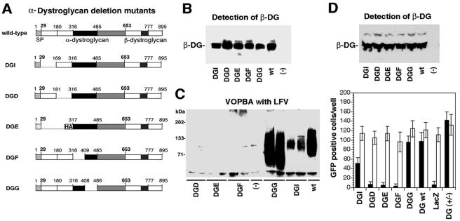
The cellular receptor for the Old World arenaviruses Lassa fever virus (LFV) and lymphocytic choriomeningitis virus (LCMV) has recently been identified as alpha-dystroglycan (alpha-DG), a cell surface receptor that provides a molecular link between the extracellular matrix and the actin-based cytoskeleton. In the present study, we show that LFV binds to alpha-DG with high affinity in the low-nanomolar range. Recombinant vesicular stomatitis virus pseudotyped with LFV glycoprotein (GP) adopted the receptor binding characteristics of LFV and depended on alpha-DG for infection of cells. Mapping of the binding site of LFV on alpha-DG revealed that LFV binding required the same domains of alpha-DG that are involved in the binding of LCMV. Further, LFV was found to efficiently compete with laminin alpha1 and alpha2 chains for alpha-DG binding. Together with our previous studies on receptor binding of the prototypic immunosuppressive LCMV isolate LCMV clone 13, these findings indicate a high degree of conservation in the receptor binding characteristics between the highly human-pathogenic LFV and murine-immunosuppressive LCMV isolates.


Figures

FIG. 1.
FIG. 1.
LFV binds α-DG with high affinity. (A) Detection of viral GPs in purified LFV and LCMV. Purified LFV strain Josiah (inactivated by γ-irradiation) and LCMV cl-13 (107 PFU/ml each) were solubilized in SDS-PAGE buffer, and 40 μl (lanes 1) and 10 μl (lanes 2) of the samples were separated by SDS-PAGE and transferred to nitrocellulose. Blots were probed with MAb 83.6 anti-LCMVGP2, using a peroxidase-conjugated secondary antibody and enhanced chemiluminescence (ECL) for detection. Molecular masses are indicated. (B) Comparison of the binding affinities for α-DG of LFV and LCMV cl-13 by VOPBA. α-DG, purified from rabbit skeletal muscle, was diluted (1, 0.1, 0.01, 0.001 μg) and blotted to nitrocellulose. LFV and LCMV cl-13 were applied at the same concentration (107 PFU/ml). Bound virus was detected by monoclonal antibodies 33.6 and 86.6 that recognize conserved epitopes of LFV and LCMV GP2, using a peroxidase-conjugated anti-mouse IgG secondary antibody and enhanced chemiluminescence (ECL). (C) Binding of α-DG to LFV, LCMV cl-13, and LCMV WE2.2. LFV Josiah (circles), LCMV cl-13 (squares), and LCMV WE2.2 (triangles) were immobilized in a microtiter plate and incubated with biotinylated α-DG. Bound biotinylated or α-DG was detected with peroxidase-conjugated streptavidin in a color reaction using ABTS substrate. OD405 was recorded in an ELISA reader. For the determination of specific binding, background binding to BSA was subtracted (mean ± standard deviation; n = 3).
FIG. 2.
FIG. 2.
Characterization of recombinant LFVGP. (A) Expression of recombinant LFVGP. HeLa cells were transfected with the expression construct pC-LFVGP (lane 1), pC-EGFP (lane 2), and empty vector (lane 3) using Lipofectamine. Two days after transfection, cells were lysed in SDS-PAGE sample buffer. As a positive control, a lysate of purified LFV virions (107 PFU/ml) was used (lane 4). Proteins were separated by SDS-PAGE, transferred to nitrocellulose, and analyzed by Western blot using MAb 83.6 anti-LCMVGP2. Bound primary antibody was detected with a peroxidase-conjugated secondary antibody using ECL for detection. Molecular masses are indicated. (B) Cell surface labeling of recombinant LFVGP. HeLa cells were transfected with pC-LFVGP (LFVGP) or empty vector (mock) as in panel A. Cell surface LFVGP was detected with MAb 83.6 anti-LCMVGP2 and a PE-conjugated secondary antibody to mouse IgG. For flow cytometry, a FACSCalibur flow cytometer was used. In histograms, the y axis represents cell numbers and the x axis represents fluorescence intensity (FL2-H for anti-mouse IgG-PE conjugate).
FIG. 3.
FIG. 3.
Recombinant vesicular stomatitis virus pseudotyped with LFVGP adopts the receptor binding characteristics of LFV and depends on α-DG for infection. (A) Blocking of VSVΔG*-LFVGP infection by γ-inactivated LFV. HeLa cells cultured in 96-well plates were blocked with the indicated PFU/cell of γ-inactivated LFV or γ-inactivated Amapari. After incubation for two hours at 4°C, cells were infected with 200 IU of VSVΔG*-LFVGP (black bars) or VSVΔG*-VSVGP (white bars). Infection was assessed after 24 h by detection of the EGFP reporter in fluorescence microscopy. Data are EGFP-positive cells per well (n = 3; ± standard deviation). (B) Infection with LFV-PS depends on α-DG. DG−/− ES cells were infected with AdV vectors containing either wild-type DG or the deletion mutant DGH at an MOI of 10. After 48 h, AdV-infected cells as well as DG−/− and DG+/− ES cells cultured in parallel were infected with 200 IU of VSVΔG*-LFVGP or VSVΔG*-VSVGP and infection was assessed after 24 h as in panel B (n = 6; ± standard deviation).
FIG. 4.
FIG. 4.
Structures within amino acids 169 to 408 are required for LFV binding. (A) Schematic representation of the α-DG deletion mutants: The putative N-terminal subdomains (white), the mucin-type domain (black), and the C-terminal globular domain (gray) of α-DG are indicated. Amino acids 653 to 895 represent β-DG with the transmembrane domain (dark box). The influenza virus hemagglutinin (HA) epitope in DGE is indicated. (B) Expression of the α-DG deletion mutants. DG−/− ES cells were infected with AdV vectors containing the α-DG variants or green fluorescent protein (−). After 48 h, cells were lysed and samples subjected to jacalin affinity chromatography. Jacalin-bound glycoproteins were separated by SDS-PAGE, transferred to nitrocellulose, and probed with anti-β-DG. wt, wild type. (C) Binding of LCMV cl-13 and LFV to wild-type and deletion mutants of α-DG. Jacalin-bound glycoproteins (B) were subjected to VOPBA with 107 PFU/ml of LFV using MAb 83.6, anti-LCMVGP2, and enhanced chemiluminescence (ECL) for detection. (D) Reconstitution of LFV-PS infectivity in DG−/− ES cells by wild-type and deletion mutants of α-DG. DG−/− ES cells were infected with AdV vectors containing wild-type and deletion mutants of α-DG or a β-galactosidase reporter gene (LacZ) at an MOI of 10. After 48 h, transgene expression was verified by detection of β-DG in total cell protein in the Western blot. Cells cultured in parallel were infected with 200 IU (MOI = 0.01) of VSVΔG*-LFVGP (black bars) or VSVΔG*-VSVGP (white bars), and infection was assessed after 24 h as in panel B. Infection levels were assessed 24 h later by the detection of GFP-positive cells by immunofluorescence (n = 6; ± standard deviation).
FIG. 5.
FIG. 5.
Amino acids 169 to 408 of α-DG contain a high-affinity binding site for LFV and LCMV. (A) Schematic representation of the α-DG fragments fused to human IgG Fc. The putative domains of α-DG are depicted as in Fig. 4, and human IgG1Fc is indicated. wt, wild type. (B) Purified α-DG-Fc fusion proteins. The α-DG-Fc fusion proteins DGFc1 through -5 were expressed in HEK293T cells and purified by protein A affinity chromatography from cell lysates. Purified proteins were analyzed by SDS-PAGE with Coomassie blue staining (B) and by Western blot using an anti-human IgG Fc antibody (C). (D) Binding of LFV to the α-DG-Fc fusion proteins. Equal amounts of purified DGFc1 through -5 were immobilized in microtiter plates and incubated with 107 PFU/ml γ-inactivated LFV. Bound virus was detected with MAb 83.6 (anti-LCMVGP2) and a peroxidase-conjugated secondary antibody in a color reaction using ABTS substrate. OD405 was recorded in an ELISA reader. For the determination of specific binding, background binding to BSA was subtracted (mean ± standard deviation; n = 3).
FIG. 6.
FIG. 6.
LFV and LCMV cl-13 compete with laminin α1 and α2 chains for α-DG binding. (A) Blocking of virus binding to α-DG by laminin-1 and laminin-2. α-DG purified from rabbit skeletal muscle was immobilized in microtiter plates and blocked with the indicated concentrations of laminin-1 (black), laminin-2 (gray), and fibronectin (white) for 4 h at room temperature. Inactivated LFV and LCMV cl-13 were added in a dilution of 107 PFU/ml containing the blocking proteins in the concentrations mentioned above for 12 h at 6°C. Bound virus was detected as in Fig. 5D. For the determination of specific binding, background binding to BSA was subtracted (mean ± standard deviation; n = 3). (B) Displacement of LFV bound to α-DG with soluble laminin-1, laminin-2, and fibronectin: Inactivated LFV and LCMV cl-13 (107 PFU/ml) were bound to immobilized α-DG. Plates were washed, and the indicated concentrations of ECM proteins were then added for 4 h at room temperature. After three wash steps, bound virus was detected as described in panel A.

References

    1. Ahmed, R., A. Salmi, L. D. Butler, J. M. Chiller, and M. B. Oldstone. 1984. Selection of genetic variants of lymphocytic choriomeningitis virus in spleens of persistently infected mice. Role in suppression of cytotoxic T lymphocyte response and viral persistence. J. Exp. Med. 160:521-540. - PMC - PubMed
    1. Baize, S., J. Kaplon, C. Faure, D. Pannetier, M. C. Georges-Courbot, and V. Deubel. 2004. Lassa virus infection of human dendritic cells and macrophages is productive but fails to activate cells. J. Immunol. 172:2861-2869. - PubMed
    1. Belkin, A. M., and K. Burridge. 1995. Association of aciculin with dystrophin and utrophin. J. Biol. Chem. 270:6328-6337. - PubMed
    1. Belkin, A. M., and K. Burridge. 1995. Localization of utrophin and aciculin at sites of cell-matrix and cell-cell adhesion in cultured cells. Exp. Cell Res. 221:132-140. - PubMed
    1. Belkin, A. M., and N. R. Smalheiser. 1996. Localization of cranin (dystroglycan) at sites of cell-matrix and cell-cell contact: recruitment to focal adhesions is dependent upon extracellular ligands. Cell Adhes. Commun. 4:281-296. - PubMed

Publication types

LinkOut - more resources