Comparative Study
. 2005 May;79(10):6005-22.
doi: 10.1128/JVI.79.10.6005-6022.2005.

Targeting human glioblastoma cells: comparison of nine viruses with oncolytic potential

Affiliations
Comparative Study

Targeting human glioblastoma cells: comparison of nine viruses with oncolytic potential

Guido Wollmann et al. J Virol. 2005 May.

Abstract

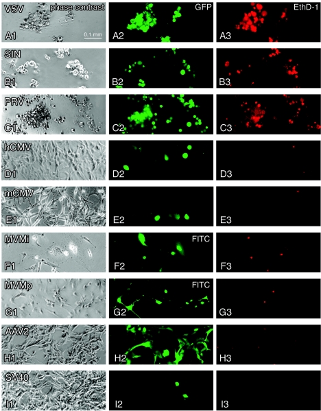
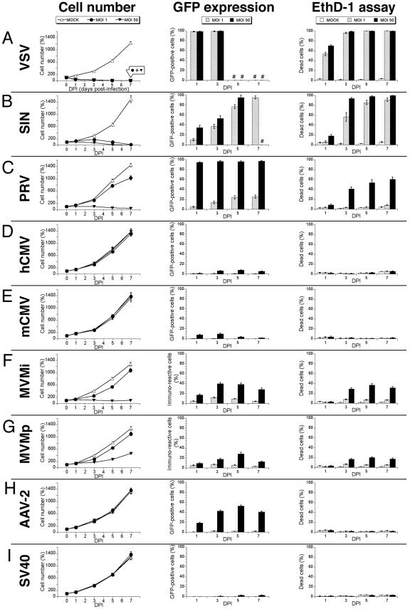
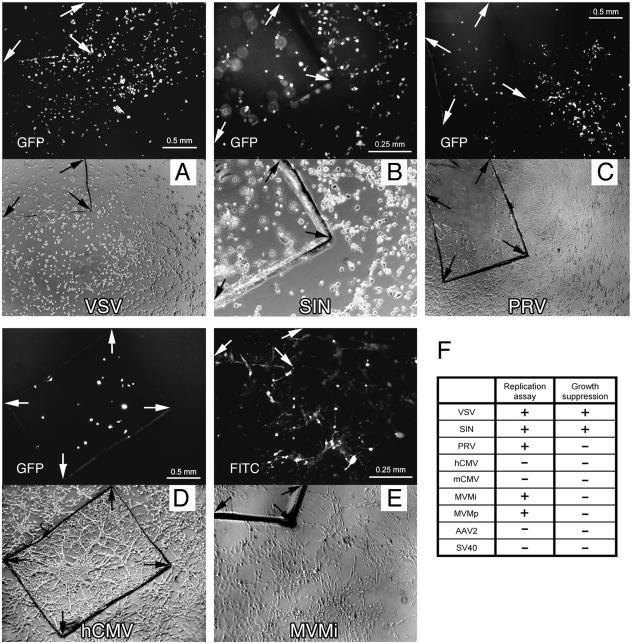
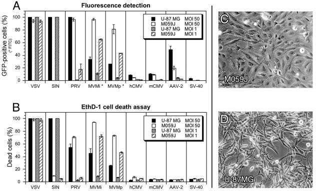
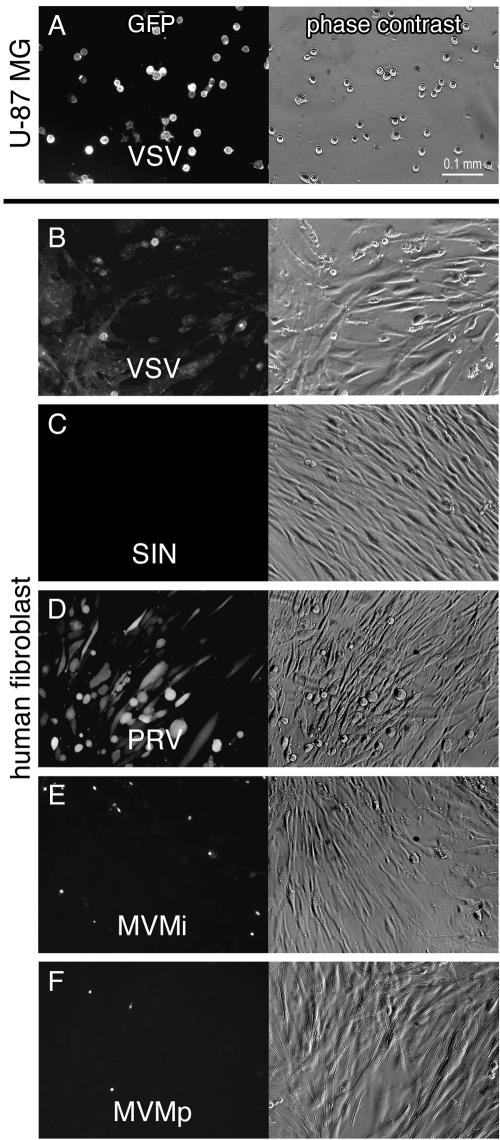
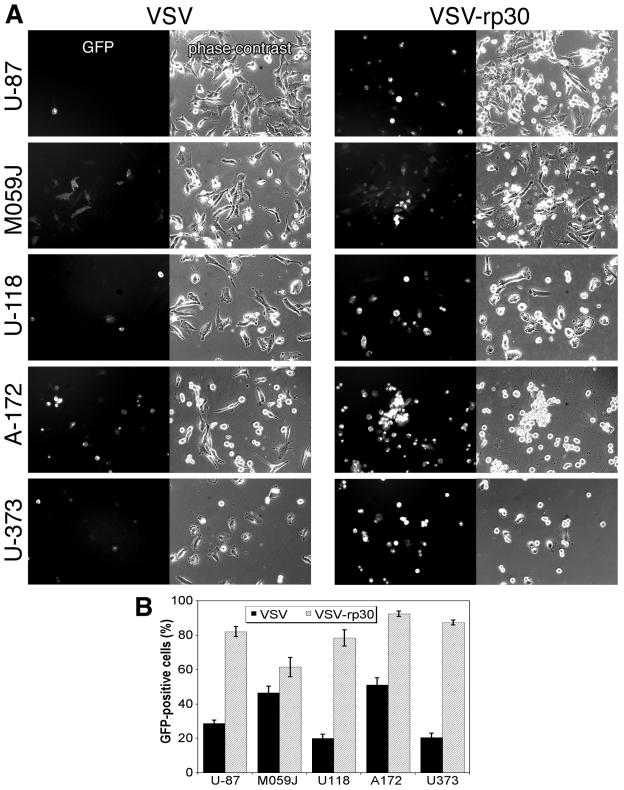
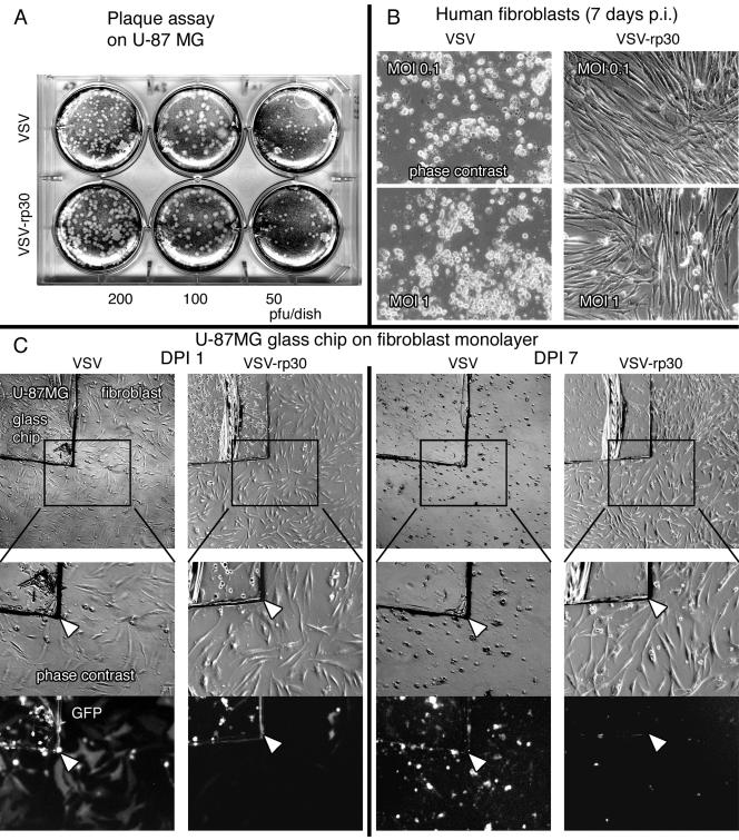
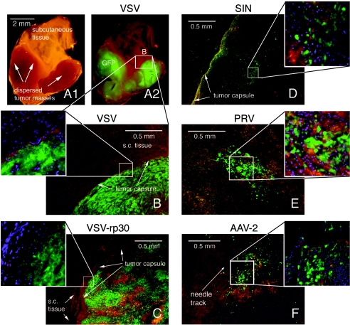
Brain tumors classified as glioblastomas have proven refractory to treatment and generally result in death within a year of diagnosis. We used seven in vitro tests and one in vivo trial to compare the efficacy of nine different viruses for targeting human glioblastoma. Green fluorescent protein (GFP)-expressing vesicular stomatitis (VSV), Sindbis virus, pseudorabies virus (PRV), adeno-associated virus (AAV), and minute virus of mice i-strain (MVMi) and MVMp all infected glioblastoma cells. Mouse and human cytomegalovirus, and simian virus 40 showed only low levels of infection or GFP expression. VSV and Sindbis virus showed strong cytolytic actions and high rates of replication and spread, leading to an elimination of glioblastoma. PRV and both MVM strains generated more modest lytic effects and replication capacity. VSV showed a similar oncolytic profile on U-87 MG and M059J glioblastoma. In contrast, Sindbis virus showed strong preference for U-87 MG, whereas MVMi and MVMp preferred M059J. Sindbis virus and both MVM strains showed highly tumor-selective actions in glioblastoma plus fibroblast coculture. VSV and Sindbis virus were serially passaged on glioblastoma cells; we isolated a variant, VSV-rp30, that had increased selectivity and lytic capacity in glioblastoma cells. VSV and Sindbis virus were very effective at replicating, spreading within, and selectively killing human glioblastoma in an in vivo mouse model, whereas PRV and AAV remained at the injection site with minimal spread. Together, these data suggest that four (VSV, Sindbis virus, MVMi, and MVMp) of the nine viruses studied merit further analysis for potential therapeutic actions on glioblastoma.


Figures

FIG. 1.
FIG. 1.
Virus infection, transgene expression, and cytolytic action in human glioblastoma cells. Representative effects of seven recombinant GFP-expressing viruses and two strains of MVM are depicted in this panel showing phase-contrast (first column), green GFP fluorescence (second column), and EthD-1 cell death staining (third column) in corresponding visual fields after U-87 MG inoculation. All cultures were infected at an initial MOI of 50. (A) Infection with VSV after 24 h. (B) SIN infection at 3 dpi. (C) PRV infection at 7 dpi. (D) Infection with human CMV at 5 dpi. A small number of cells accumulate high levels of GFP and show CPEs. (E) Infection with mouse CMV at 3 dpi. Green cells appeared mostly with normal morphology. The cellular GFP expression was weak. (F) MVMi infection at 3 dpi. Immunocytochemistry revealed predominantly nuclear staining of viral proteins NS1/NS2. (G) MVMp infection at 5 dpi. (H) Infection with AAV2 at 5 dpi. (I) Infection with SV40 at 7 dpi.
FIG.2.
FIG.2.
Growth curves, fluorescence detection, and cell death analysis. U-87 MG cells were infected at an MOI of 1 or 50 or not infected (MOCK). The data are presented as means ± the standard errors of the mean. (A) VSV rapidly infected and killed glioblastoma cells regardless of the initial viral concentration, leading to nearly identical growth curve suppression by both MOIs. #, Lack of any viable cells. (B) Upon SIN infection tumor cells stop dividing and ultimately died. Some dead cells might have lost their cytosolic GFP through diffusion explaining the higher EthD-1 values. (C) PRV exerts a tumor-suppressive effect at a higher MOI. (D and E) Human (D) or mouse (E) CMV infection did not affect tumor cell growth. (F) MVMi infection stops tumor cell growth at a high MOI. (G) MVMp infection at MOI 50 moderately slows down tumor progression. (H) AAV2 showed considerable infectivity at MOI 50 without disturbing cell viability. (I) Very low infection rates of SV40 at MOI 50.
FIG. 3.
FIG. 3.
Virus replication and spread. Small glass chips carrying infected U-87 MG cells were washed and transferred onto a native U-87 MG cell layer. Each set of photomicrographs depicts corresponding fields of fluorescence and phase contrast. (A) Widespread GFP expression after VSV infection in the culture dish at 1 dpi. (B) Replication of SIN was observed with a 3-day latency, inflicting extensive cell death throughout the culture. Due to the different plane, cells located on the glass chip are out of focus. (C) PRV spread to close areas in a fanning-out fashion, indicating a potential cell-to-cell distribution. The integrity of the monolayer was not affected. (D) Human CMV did not replicate and spread. (E) MVMi immunopositive cells were detected at 3 dpi in the area fringing the glass chip. At later time points, groups of cells remote from the glass chip stained positive for MVM infection. (F) Table summarizing virus replication and the effect on the growth of U-87 MG monolayer.
FIG. 4.
FIG. 4.
Generalization in different human glioblastoma lineages of virus infectivity and cytolysis. Two different glioblastoma cell lines were compared for viral infection and associated cytotoxicity. Analysis was carried out at 3 dpi for VSV and at 5 dpi for the other viruses. The data represent means ± the standard errors of the mean. (A) Considerable differences in tropism were observed with SIN, MVMi, and MVMp and to a smaller extent with PRV and AAV. (B) Different cytolytic potential of SIN and both MVM strains showed strong correlation to infectivity. (C) Photomicrograph depicts the typical cell morphology of M059J cells in 5-day-old cultures. (D) Typical image of U-87 MG cells after 5 days in culture.
FIG. 5.
FIG. 5.
Infection of cocultured human glioblastoma and control fibroblasts. Images show phase-contrast and GFP expression or fluorescein isothiocyanate (FITC) staining of human fibroblasts cocultured withU-87 MG cells in the same dish. The virus concentration was MOI 1. (A) At 24 h after VSV infection, all tumor cells showed GFP expression and CPEs. (B) In the same dish, some fibroblasts showed GFP expression. (C) SIN infection at 3 dpi, with no signs of infection among the fibroblast cells. (D) Preferential infection of human fibroblasts by PRV at 3 dpi. (E and F) Few signs of infection with MVMi and MVMp among human fibroblasts. Images were taken after immunocytochemistry at 3 dpi.
FIG. 6.
FIG. 6.
Glioblastoma-adapted VSV-rp30: enhanced targeting of diverse glioma phenotypes. The glioblastoma-adapted strain VSV-rp30 was compared to the original VSV-G/GFP. (A) VSV-rp30 presents a strong tropism for numerous tested glioblastoma cell lines. Representative photomicrographs are shown here for U-87 MG, M059J, U-118 MG, A-172, and U-373 MG cells. The MOI for VSV and VSV-rp30 was 10. (B) Bar graph comparing the infection rates of VSV and VSV-rp30 on U-373, A-172, U-118, M059J, and U-87 at 10 MOI. Values represent mean of 10 microscopic fields. In each cell line, pairs of micrographs showing VSV or VSV-rp30 and analysis of cell infection were made at the same time postinoculation (<12 h).
FIG. 7.
FIG. 7.
Glioblastoma-adapted VSV-rp30: attenuation of control cell infection. (A) Virus suspensions were tested with plaque assays prior to each experiment to ensure equal viral load for this comparative study. (B) VSV-rp30 was considerably attenuated for infection and cytotoxicity of normal human fibroblasts compared to the original VSV. (C) Coculture infection. Tumor cells were grown on a glass chip and transferred onto a fibroblast layer prior to adding either VSV-G/GFP or VSV-rp30 at an MOI of 1. The photomicrographs illustrate patterns of GFP expression and presence of cytopathic effects at 24 hpi (two left columns) and the long-term outcome at 7 dpi (two right columns).
FIG. 8.
FIG. 8.
Virus infection of glioblastoma xenografts in vivo. Images of tumor sections were taken using a multichannel fluorescence filter. Green indicates GFP expression, blue indicates nuclei stained with DAPI, and red-brown areas mark tissue background. (A1) Macroscopic aspect of a tissue block containing two small tumor masses that were remote from the original site of VSV injection. (A2) At 10 dpi, VSV selectively infected the small tumor masses. (B) Microscopic image of tumor mass shown in A. Note the demarcation at the tumor capsule. (C) At 10 days after intratumoral injection, VSV-rp30 has infected the whole tumor mass without crossing the tumor border (inset). (D) Intratumoral injection of SIN led to multiple widespread green islands and a distinct subcapsular green fluorescent band, indicating active viral spread. (E) Intratumoral injection with PRV at 10 dpi. The central area of the tumor is depicted. A similar spread was seen at 3 dpi. (F) Diffusion control with replication deficient AAV2. GFP expression was restricted to a 0.5-mm radius around the needle track.

References

    1. Ali-Osman, F. 1999. Brain tumors, p. 167-184. In J. R. W. Masters and B. Palson (ed.), Human cell culture, vol. 2. Kluwer Academic Publishers, New York, N.Y.
    1. American Cancer Society Surveillance Research. 2002. Cancer facts and figures 2002. American Cancer Society, Inc., Atlanta, Ga.
    1. Anderson, C. W., and M. J. Allalunis-Turner. 2000. Human TP53 from the malignant glioma-derived cell lines M059J and M059K has a cancer-associated mutation in exon 8. Radiat. Res. 154:473-476. - PubMed
    1. Anderson, C. W., J. J. Dunn, P. I. Freimuth, A. M. Galloway, and M. J. Allalunis-Turner. 2001. Frameshift mutation in PRKDC, the gene for DNA-PKcs, in the DNA repair-defective, human, glioma-derived cell line M059J. Radiat. Res. 156:2-9. - PubMed
    1. Balachandran, S., and G. N. Barber. 2000. Vesicular stomatitis virus (VSV) therapy of tumors. IUBMB Life 50:135-138. - PubMed

Publication types