Comparative Study
. 2005 May;79(10):6152-61.
doi: 10.1128/JVI.79.10.6152-6161.2005.

Cellular localization and antigenic characterization of crimean-congo hemorrhagic fever virus glycoproteins

Affiliations
Comparative Study

Cellular localization and antigenic characterization of crimean-congo hemorrhagic fever virus glycoproteins

Andrea Bertolotti-Ciarlet et al. J Virol. 2005 May.

Abstract

Crimean-Congo hemorrhagic fever virus (CCHFV), a member of the genus Nairovirus of the family Bunyaviridae, causes severe disease with high rates of mortality in humans. The CCHFV M RNA segment encodes the virus glycoproteins G(N) and G(C). To understand the processing and intracellular localization of the CCHFV glycoproteins as well as their neutralization and protection determinants, we produced and characterized monoclonal antibodies (MAbs) specific for both G(N) and G(C). Using these MAbs, we found that G(N) predominantly colocalized with a Golgi marker when expressed alone or with G(C), while G(C) was transported to the Golgi apparatus only in the presence of G(N). Both proteins remained endo-beta-N-acetylglucosaminidase H sensitive, indicating that the CCHFV glycoproteins are most likely targeted to the cis Golgi apparatus. Golgi targeting information partly resides within the G(N) ectodomain, because a soluble version of G(N) lacking its transmembrane and cytoplasmic domains also localized to the Golgi apparatus. Coexpression of soluble versions of G(N) and G(C) also resulted in localization of soluble G(C) to the Golgi apparatus, indicating that the ectodomains of these proteins are sufficient for the interactions needed for Golgi targeting. Finally, the mucin-like and P35 domains, located at the N terminus of the G(N) precursor protein and removed posttranslationally by endoproteolysis, were required for Golgi targeting of G(N) when it was expressed alone but were dispensable when G(C) was coexpressed. In neutralization assays on SW-13 cells, MAbs to G(C), but not to G(N), prevented CCHFV infection. However, only a subset of G(C) MAbs protected mice in passive-immunization experiments, while some nonneutralizing G(N) MAbs efficiently protected animals from a lethal CCHFV challenge. Thus, neutralization of CCHFV likely depends not only on the properties of the antibody, but on host cell factors as well. In addition, nonneutralizing antibody-dependent mechanisms, such as antibody-dependent cell-mediated cytotoxicity, may be involved in the in vivo protection seen with the MAbs to G(C).


Figures

FIG. 1.
FIG. 1.
Schematic representation of CCHFV M segment constructs. Predicted N-linked glycosylation sites are indicated with “Y.” The filled triangles (▴) indicate the predicted conserved cleavage sites with the four conserved amino acid cleavage sites indicated (59), and the black bars indicate the predicted TMs. Specific constructs used in this study are also indicated by black lines, indicating which portions of the CCHFV M segment are included in each construct. The gray boxes on the constructs GN-RSKR and GN-RRLL indicate the inclusion of the hen egg white lysozyme signal peptide at the N terminus of the protein (6). A V5 epitope tag was positioned at the C terminus of each construct; these tags are represented as black boxes on the far-right side of each construct. Finally, the amino acids included in each construct are indicated on the right column, using the first methionine in the IbAr10200 strain as residue 1.
FIG. 2.
FIG. 2.
Analysis of GN and GC cellular localization. HeLa cells were transfected with a pCAGGS vector expressing the full-length M segment of CCHFV. After 24 h, the cells were fixed and stained using the indicated MAbs in addition to an antibody specific for the Golgi resident protein TGN46 as described in Materials and Methods. The images show the Golgi apparatus (red) and the glycoproteins (green) merged. Yellow indicates colocalization. The secondary antibodies used were Alexa Fluor 488 (goat anti-mouse) and Alexa Fluor 594 (goat anti-sheep). Magnification, ×60; bar = 100 nm.
FIG. 3.
FIG. 3.
MAb 11E7 directed against GC recognizes a linear epitope. HEK 293T cells were transfected with the pCAGGS vector containing the entire CCHFV M segment (M), infected with vaccinia virus vTF1.1 and transfected with the pDNA3.1 constructs containing the GN or GC full-length gene with a V5 tag at the C terminus, or mock transfected. Alternatively, SW-13 cells infected with CCHFV were lysed and analyzed by Western blotting (lane labeled “virus”). Western blotting was performed utilizing MAb 11E7. Molecular size markers (in kilodaltons) are noted at the right of each blot.
FIG. 4.
FIG. 4.
Correlation between in vitro plaque reduction and in vivo protection. Passive immunization with CCHFV MAb against GC (A) or against GN (B). The percentage of protection represents the proportion of animals that survived challenge with respect to the total number of animals treated. Protection was determined in 2- to 3-day-old mice as described in Materials and Methods. The antibodies were provided 24 h before or 24 h after intraperitoneal challenge with the IbAr10200 strain of CCHFV. The plaque reduction neutralization titers are shown at the bottom of each plot for each antibody and represent 80% plaque reduction in SW-13 monolayer cells.
FIG. 5.
FIG. 5.
(A) Localization of CCHFV glycoproteins expressed independently. HeLa cells were infected with recombinant vaccinia virus vTF1.1 and transfected with GN-FL (a), GC-FL (b), or both constructs (c). A schematic of the constructs is shown in Fig. 1. Twenty hours posttransfection, the cells were analyzed for the localization of the proteins by IF using MAbs specific for GN (a) or GC (b and c). The images show the glycoproteins (in green), the Golgi apparatus (in red), and the extent of colocalization (in yellow). Bar = 100 nm. (B) Endo-H treatment of CCHFV glycoproteins. HEK 293T cells were transfected with the GN-FL, GC-FL, or GN-RRLL constructs separately or together in the presence of recombinant vaccinia virus-expressing T7 polymerase. The lysates were treated with PNGaseF (F) or endo-H (H) or were not treated (N). The lysates were analyzed by Western blotting with mouse anti-V5 antibody. Molecular size markers (in kilodaltons) are noted at the left.
FIG. 6.
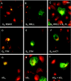
FIG. 6.
Immunofluorescence analysis of the CCHFV glycoprotein constructs. HeLa cells were transfected with the indicated CCHFV constructs (shown schematically in Fig. 1) in the presence of recombinant T7 RNA polymerase expressed by vaccinia virus vTF1.1. Twenty hours posttransfection, the cells were fixed and stained using MAb 11E7 against GC (panels H and I) and MAb 8F10 against GN (panels A through G) (green) and the TGN46 antibody for Golgi localization (red). Nuclear staining is shown in blue. Bar = 100 nm.

References

    1. Alexander, W. A., B. Moss, and T. R. Fuerst. 1992. Regulated expression of foreign genes in vaccinia virus under the control of bacteriophage T7 RNA polymerase and the Escherichia coli lac repressor. J. Virol. 66:2934-2942. - PMC - PubMed
    1. Anderson, G. W., Jr., and J. F. Smith. 1987. Immunoelectron microscopy of Rift Valley fever viral morphogenesis in primary rat hepatocytes. Virology 161:91-100. - PubMed
    1. Andersson, A. M., L. Melin, A. Bean, and R. F. Pettersson. 1997. A retention signal necessary and sufficient for Golgi localization maps to the cytoplasmic tail of a Bunyaviridae (Uukuniemi virus) membrane glycoprotein. J. Virol. 71:4717-4727. - PMC - PubMed
    1. Andersson, A. M., L. Melin, R. Persson, E. Raschperger, L. Wikström, and R. F. Pettersson. 1997. Processing and membrane topology of the spike proteins G1 and G2 of Uukuniemi virus. J. Virol. 71:218-225. - PMC - PubMed
    1. Andersson, A. M., and R. F. Pettersson. 1998. Targeting of a short peptide derived from the cytoplasmic tail of the G1 membrane glycoprotein of Uukuniemi virus (Bunyaviridae) to the Golgi complex. J. Virol. 72:9585-9596. - PMC - PubMed

Publication types

MeSH terms