. 2005 May;79(10):6194-206.
doi: 10.1128/JVI.79.10.6194-6206.2005.

Carboxyl-proximal regions of reovirus nonstructural protein muNS necessary and sufficient for forming factory-like inclusions

Affiliations

Carboxyl-proximal regions of reovirus nonstructural protein muNS necessary and sufficient for forming factory-like inclusions

Teresa J Broering et al. J Virol. 2005 May.

Abstract

Mammalian orthoreoviruses are believed to replicate in distinctive, cytoplasmic inclusion bodies, commonly called viral factories or viroplasms. The viral nonstructural protein muNS has been implicated in forming the matrix of these structures, as well as in recruiting other components to them for putative roles in genome replication and particle assembly. In this study, we sought to identify the regions of muNS that are involved in forming factory-like inclusions in transfected cells in the absence of infection or other viral proteins. Sequences in the carboxyl-terminal one-third of the 721-residue muNS protein were linked to this activity. Deletion of as few as eight residues from the carboxyl terminus of muNS resulted in loss of inclusion formation, suggesting that some portion of these residues is required for the phenotype. A region spanning residues 471 to 721 of muNS was the smallest one shown to be sufficient for forming factory-like inclusions. The region from positions 471 to 721 (471-721 region) includes both of two previously predicted coiled-coil segments in muNS, suggesting that one or both of these segments may also be required for inclusion formation. Deletion of the more amino-terminal one of the two predicted coiled-coil segments from the 471-721 region resulted in loss of the phenotype, although replacement of this segment with Aequorea victoria green fluorescent protein, which is known to weakly dimerize, largely restored inclusion formation. Sequences between the two predicted coiled-coil segments were also required for forming factory-like inclusions, and mutation of either one His residue (His570) or one Cys residue (Cys572) within these sequences disrupted the phenotype. The His and Cys residues are part of a small consensus motif that is conserved across muNS homologs from avian orthoreoviruses and aquareoviruses, suggesting this motif may have a common function in these related viruses. The inclusion-forming 471-721 region of muNS was shown to provide a useful platform for the presentation of peptides for studies of protein-protein association through colocalization to factory-like inclusions in transfected cells.


Figures
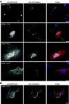
FIG. 1.
FIG. 1.
IF microscopy of C- terminally truncated μNS proteins. CV-1 cells were transfected with plasmids to express the indicated proteins. The cells were then fixed at 18 h p.t. for subsequent immunostaining. Nuclei were counterstained with DAPI (blue). Scale bars, 10 μm. (A) The cells were immunostained both with rabbit anti-μNS IgG conjugated to Texas red (left column, red in right column) and with mouse MAb FK2 for conjugated ubiquitin (cUb), followed by goat anti-mouse IgG conjugated to Alexa 488 (center column, green in right column). (B) The cells were immunostained both with rabbit anti-μNS IgG followed by goat anti-rabbit IgG conjugated to Alexa 594 (left column, red in right column) and with rabbit anti-μ2 IgG conjugated to Alexa 488 (center column, green in right column).
FIG. 2.
FIG. 2.
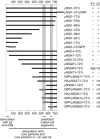
Summary of μNS truncations and their activities. The full-length μNS protein is indicated by a horizontally elongated black bar spanning residues 1 to 721 (positions numbered above and below). The μNS truncation mutants are also shown as black bars spanning the approximate portion of μNS that each represents. Enhanced A. victoria GFP fused to the N or C terminus of μNS in some cases is represented by an open bar. An influenza virus HA epitope fused to the N terminus of μNS in some cases is represented by an open circle. Approximate extents of the coiled-coil segments predicted by Multicoil are indicated by vertically elongated gray bars. The capacity of each protein to form factory-like inclusions in transfected cells (Inc) and to colocalize with T1L μ2 in transfected cells (μ2) is indicated as positive (+), negative (−), or not determined (nd). Localized structures that costained for conjugated ubiquitin were concluded to be aggregates of misfolded protein (agg). The results for μNS(1-721), μNS(1-721)/GFP, μNS(1-41)/GFP, μNS(14-721), and μNS(41-721) have been reported previously (7, 26).
FIG. 3.
FIG. 3.
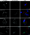
IF microscopy of N-terminally truncated μNS proteins. CV-1 cells were transfected with plasmids to express the indicated proteins. The cells were then fixed and stained as described for Fig. 1A. Scale bars, 10 μm.
FIG. 4.
FIG. 4.
Immunoblotting and IF microscopy of GFP-tagged derivatives of μNS. CV-1 cells were transfected with plasmids to express GFP or μNS-GFP fusions as indicated and then analyzed at 18 h p.t. by immunoblotting (A) or IF microscopy (B). Scale bars, 10 μm. (A) Whole-cell lysates were separated by SDS-PAGE, transferred to nitrocellulose, and immunoblotted with mouse MAb JL8 for GFP followed by goat anti-mouse IgG conjugated to alkaline phosphatase. Positions of molecular weight markers are indicated (in kilodaltons) to the left of each panel. (B) After fixation, cells were immunostained with mouse MAb JL8 for GFP, followed by goat anti-mouse IgG conjugated to Alexa 488.
FIG. 5.
FIG. 5.
IF microscopy of HA-tagged or untagged versions of μNS(561-721) and μNS(471-721). CV-1 cells were transfected with plasmids to express the proteins as indicated. The cells were then fixed at 18 h p.t. and immunostained either with mouse MAb HA.11 for the influenza virus HA epitope, followed by goat anti-mouse IgG conjugated to Alexa 488 (left panels), or with rabbit anti-μNS IgG, followed by goat anti-rabbit IgG conjugated to Alexa 594 (right panels). Scale bars, 10 μm.
FIG. 6.
FIG. 6.
Sequence conservation in the “linker” region of μNS homologs and IF microscopy of μNS proteins with mutations at His and/or Cys residues in the consensus motif. (A) Sequence conservation. Sequences are shown in single-letter code, with position numbers indicated at left and right. The consensus motif was defined by comparing the illustrated region from the μNS homologs of 3 mammalian orthoreoviruses (mORV), 12 avian orthoreoviruses (aORV) (all identical in this region), and 2 aquareoviruses (AqRV) (both identical in this region). Conserved positions are highlighted by being boxed. Asterisks indicate conserved positions corresponding to His570 and Cys572 in mammalian orthoreovirus μNS. These and other His and Cys residues in or near the consensus motif are highlighted by being circled. (B) IF microscopy. CV-1 cells were transfected with plasmids to express the indicated proteins. The cells were then fixed at 18 h p.t. and immunostained both with rabbit anti-μNS IgG conjugated to Oregon Green and with mouse MAb FK2 for conjugated ubiquitin, followed by goat anti-mouse IgG conjugated to Alexa 594. Nuclei were counterstained with DAPI. Only the anti-μNS staining is shown since no colocalization with FK2 staining was apparent. Scale bars, 10 μm.
FIG. 7.
FIG. 7.
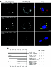
IF microscopy of σNS coexpressed with μNS-GFP fusions. (A) CV-1 cells were transfected with plasmids to express the proteins indicated. The cells were then fixed at 18 h p.t. and immunostained with σNS-specific mouse MAb 3E10, followed by goat anti-mouse IgG conjugated to Alexa 594 (center column, red in right column). GFP-containing fusions were visualized directly (left column, green in right column). Nuclei were counterstained with DAPI (blue). Scale bars, 10 μm. (B) Summary of the fusions and their activities. See Fig. 2 legend for explanations of most details. Dashed lines indicate internally deleted regions of μNS. The capacity of each protein to colocalize with T1L σNS in transfected cells (σNS) is indicated as positive (+), negative (−), or unknown (?). The results for μNS(1-721), μNS(1-721)/GFP, μNS(1-41)/GFP, μNS(14-721), and μNS(41-721) have been reported previously (7, 26).

References

    1. Antczak, J. B., and W. K. Joklik. 1992. Reovirus genome segment assortment into progeny genomes studied by the use of monoclonal antibodies directed against reovirus proteins. Virology 187:760-776. - PubMed
    1. Attoui, H., Q. Fang, F. M. Jaafar, J. F. Cantaloube, P. Biagini, P. De Micco, and X. De Lamballerie. 2002. Common evolutionary origin of aquareoviruses and orthoreoviruses revealed by genome characterization of golden shiner reovirus, grass carp reovirus, striped bass reovirus and golden ide reovirus (genus Aquareovirus, family Reoviridae). J. Gen. Virol. 83:1941-1951. - PubMed
    1. Becker, M. M., M. I. Goral, P. R. Hazelton, G. S. Baer, S. E. Rodgers, E. G. Brown, K. M. Coombs, and T. S. Dermody. 2001. Reovirus σNS protein is required for nucleation of viral assembly complexes and formation of viral inclusions. J. Virol. 75:1459-1475. - PMC - PubMed
    1. Becker, M. M., T. R. Peters, and T. S. Dermody. 2003. Reovirus σNS and μNS proteins form cytoplasmic inclusion structures in the absence of viral infection. J. Virol. 77:5948-5963. - PMC - PubMed
    1. Broering, T., A. McCutcheon, V. Centonze, and M. Nibert. 2000. Reovirus nonstructural protein μNS binds to reovirus cores but does not inhibit their transcription activity. J. Virol. 74:5516-5524. - PMC - PubMed

Publication types

Substances

LinkOut - more resources