. 2005 May;79(10):6207-15.
doi: 10.1128/JVI.79.10.6207-6215.2005.

Relocalization of the Mre11-Rad50-Nbs1 complex by the adenovirus E4 ORF3 protein is required for viral replication

Affiliations

Relocalization of the Mre11-Rad50-Nbs1 complex by the adenovirus E4 ORF3 protein is required for viral replication

Jared D Evans et al. J Virol. 2005 May.

Abstract

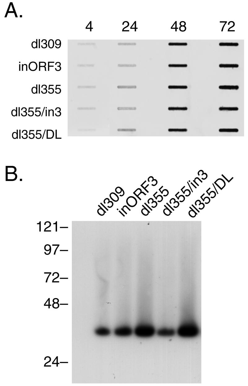
Adenovirus replication is controlled by the relocalization or modification of nuclear protein complexes, including promyelocytic leukemia protein (PML) nuclear domains and the Mre11-Rad50-Nbs1 (MRN) DNA damage machinery. In this study, we demonstrated that the E4 ORF3 protein effects the relocalization of both PML and MRN proteins to similar structures within the nucleus at early times after infection. These proteins colocalize with E4 ORF3. Through the analysis of specific viral mutants, we found a direct correlation between MRN reorganization at early times after infection and the establishment of viral DNA replication domains. Further, the reorganization of MRN components may be uncoupled from the ability of E4 ORF3 to rearrange PML. At later stages of infection, components of the MRN complex disperse within the nucleus, Nbs1 is found within viral replication centers, Rad50 remains localized with E4 ORF3, and Mre11 is degraded. The importance of viral regulation of the MRN complex is underscored by the complementation of E4 mutant viruses in cells that lack Mre11 or Nbs1 activity. These results illustrate the importance of nuclear organization in virus growth and suggest that E4 ORF3 regulates activities in both PML nuclear bodies and the MRN complex to stimulate the viral replication program.


Figures

FIG. 1.
FIG. 1.
Viral growth kinetics in A549 cells. A549 cells were infected with wild-type or mutant viruses at a multiplicity of 200 particles per cell. Virus yields in cellular lysates were measured at the times indicated by plaque assays with W162 cells and are presented as PFU per milliliter. Symbols representing the individual viruses are indicated below the growth curves. The results represent the averages of three independent experiments.
FIG. 2.
FIG. 2.
Viral DNA and protein accumulation in A549 cells. (A) A549 cells were infected with wild-type or mutant viruses at a multiplicity of 200 particles per cell. Total nuclear DNA was isolated at the times indicated and diluted (1:10, 6 h; 1:30, 12 h; 1:100, 18 h) and then applied to a nylon membrane by using a slot blot apparatus. The blot was hybridized with a fluorescently labeled probe corresponding to the left end of the Ad5 genome. The signal was detected and quantified by using a Molecular Dynamics Storm 860 PhosphorImager and ImageQuant software. (B) A549 cells were infected with E1 replacement viruses expressing HA-tagged E4 ORF3 wild-type (WT) and mutant proteins. Infected cells were harvested at 12 h postinfection and lysed in radioimmunoprecipitation buffer. Proteins were separated by SDS-polyacrylamide gel electrophoresis, transferred to a nylon membrane, probed with anti-HA mouse monoclonal antibody, and visualized by enhanced chemiluminescence.
FIG. 3.
FIG. 3.
Rearrangement of MRN components by E4 ORF3. A549 cells were infected with wild-type Ad5 (dl309) at a multiplicity of 200 particles per cell on glass coverslips. At 6 h after infection, the cells were fixed and incubated with specific antibodies against Mre11 (B), Rad50 (F), Nbs1 (J), and PML (N) (MRNP) and against E4 ORF3 (C, G, K, and O). Mre11, Rad50, Nbs1, and PML were visualized with FITC-labeled secondary antibody, and E4 ORF was visualized with TRITC-labeled secondary antibody. FITC and TRITC signals in Ad-infected cells were merged (D, H, L, and P). Uninfected (Uninf.) A549 cells were analyzed for Mre11, Rad50, Nbs1, and PML in the same manner and visualized with FITC-labeled secondary antibody (A, E, I, and M, respectively).
FIG. 4.
FIG. 4.
Localization of MRN components in comparison to Ad replication centers. A549 cells were infected with wild-type Ad5 (dl309; ORF3+/ORF6+) or dl355 (ORF3+/ORF6) at a multiplicity of 200 particles per cell on glass coverslips. At 15 h after infection, the cells were fixed and incubated with specific antibodies against Mre11 (A and M), Rad50 (E), Nbs1 (I), E4 ORF3 (B, F, J, and N), and Ad DBP (C, G, K, and O). Mre11, Rad50, and Nbs1 were visualized with FITC-labeled secondary antibody, E4 ORF3 was visualized with TRITC-labeled secondary antibody, and Ad DBP was visualized with Alexa-labeled secondary antibody. FITC, TRITC, and Alexa signals were merged (D, H, L, and P).
FIG. 5.
FIG. 5.
Rad50 relocalization with E4 ORF3 mutants. A549 cells were infected with dl355 (ORF3+/ORF6) and various E4 ORF3 mutant viruses in a dl355 background at a multiplicity of 200 particles per cell on glass coverslips. At 6 h after infection, the cells were fixed and incubated with specific antibodies against Rad50 (A, C, E, G, I, and K), PML (B, D, F, H, J, and L), and E4 ORF3 (all panels). Rad50 and PML were visualized with FITC-labeled secondary antibody, and E4 ORF3 was visualized with TRITC-labeled secondary antibody. Merges of the FITC and TRITC signals are shown in all panels. WT, wild type.
FIG. 6.
FIG. 6.
Viral DNA accumulation and concatenation in GM07166 (Nbs1) cells. (A) GM07166 cells (Nbs1) were infected with wild-type or mutant viruses at a multiplicity of 200 particles per cell. Total nuclear DNA was isolated at the times indicated and diluted (1:5, 4 h; 1:10, 24 h; 1:100, 48 h; 1:100, 72 h) and then applied to a nylon membrane by using a slot blot apparatus. The blot was hybridized with a fluorescently labeled probe corresponding to the left end of the Ad5 genome. The signal was detected and quantified by using a Molecular Dynamics Storm 860 PhosphorImager and ImageQuant software. (B) GM07166 cells were infected with wild-type or mutant viruses at a multiplicity of 200 particles per cell. Cells were harvested at 72 h postinfection, and nuclear DNA was prepared for PFGE as described in Materials and Methods. Following electrophoresis, the DNA was transferred to a nylon membrane, probed with a 32P-labeled Ad total genome probe, and visualized by autoradiography. Molecular weight standards (in kilobase pairs) are indicated on the left.

References

    1. Bischof, O., S. Kim, J. Irving, S. Beresten, N. Ellis, and J. Campisi. 2001. Regulation and localization of the Bloom syndrome protein in response to DNA damage. J. Cell Biol. 153:367-380. - PMC - PubMed
    1. Borden, K. L. 2002. Pondering the promyelocytic leukemia protein (PML) puzzle: possible functions for PML nuclear bodies. Mol. Cell. Biol. 22:5259-5269. - PMC - PubMed
    1. Boyer, J., K. Rohleder, and G. Ketner. 1999. Adenovirus E4 34k and E4 11k inhibit double strand break repair and are physically associated with the cellular DNA-dependent protein kinase. Virology 263:307-312. - PubMed
    1. Bridge, E., and G. Ketner. 1989. Redundant control of adenovirus late gene expression by early region 4. J. Virol. 63:631-638. - PMC - PubMed
    1. Carbone, R., M. Pearson, S. Minucci, and P. Pelicci. 2002. PML NBs associate with the hMre11 complex and p53 at sites of irradiation induced DNA damage. Oncogene 21:1633-1640. - PubMed

Publication types

MeSH terms