. 2005 May;79(10):6216-26.
doi: 10.1128/JVI.79.10.6216-6226.2005.

Unusual topological arrangement of structural motifs in the baboon reovirus fusion-associated small transmembrane protein

Affiliations

Unusual topological arrangement of structural motifs in the baboon reovirus fusion-associated small transmembrane protein

Sandra Dawe et al. J Virol. 2005 May.

Abstract

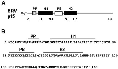
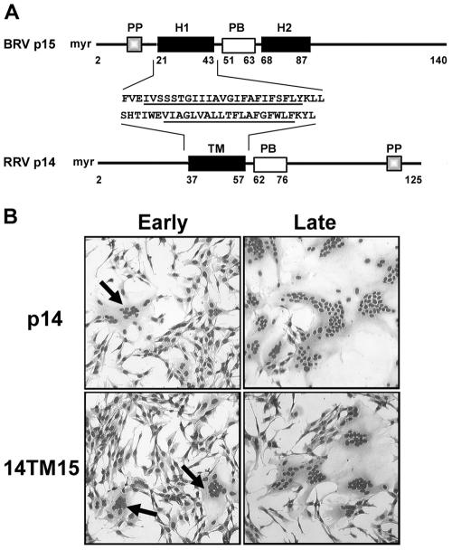
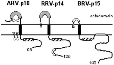
Select members of the Reoviridae are the only nonenveloped viruses known to induce syncytium formation. The fusogenic orthoreoviruses accomplish cell-cell fusion through a distinct class of membrane fusion-inducing proteins referred to as the fusion-associated small transmembrane (FAST) proteins. The p15 membrane fusion protein of baboon reovirus is unique among the FAST proteins in that it contains two hydrophobic regions (H1 and H2) recognized as potential transmembrane (TM) domains, suggesting a polytopic topology. However, detailed topological analysis of p15 indicated only the H1 domain is membrane spanning. In the absence of an N-terminal signal peptide, the H1 TM domain serves as a reverse signal-anchor to direct p15 membrane insertion and a bitopic N(exoplasmic)/C(cytoplasmic) topology. This topology results in the translocation of the smallest ectodomain ( approximately 20 residues) of any known viral fusion protein, with the majority of p15 positioned on the cytosolic side of the membrane. Mutagenic analysis indicated the unusual presence of an N-terminal myristic acid on the small p15 ectodomain is essential to the fusion process. Furthermore, the only other hydrophobic region (H2) present in p15, aside from the TM domain, is located within the endodomain. Consequently, the p15 ectodomain is devoid of a fusion peptide motif, a hallmark feature of membrane fusion proteins. The exceedingly small, myristoylated ectodomain and the unusual topological distribution of structural motifs in this nonenveloped virus membrane fusion protein necessitate alternate models of protein-mediated membrane fusion.


Figures

FIG. 1.
FIG. 1.
BRV p15 structural motifs. (A) Representation of p15 indicating the N-terminal myristate moiety (myr), the polyproline motif (PP), the two hydrophobic motifs (H1 and H2), and the polybasic motif (PB). Numbers refer to residue positions. (B) Sequence of the BRV p15 protein. The residues corresponding to the motifs presented in panel A are overlined.
FIG. 2.
FIG. 2.
BRV p15 is targeted to the endoplasmic reticulum. (A) Transfected cells expressing p15 were incubated in the absence or presence of brefeldin A, as outlined in Materials and Methods. The extent of syncytium formation was determined by Wright-Giemsa staining to reveal the presence of clustered nuclei in polykaryons, and images were captured by light microscopy at ×170 magnification. (B) Transfected cells expressing p15 were methanol-fixed and coimmunostained using rabbit anti-p15 polyclonal antiserum and FITC-conjugated goat anti-rabbit F(ab′)2 (α-p15 panel), followed by mouse anti-calnexin and rhodamine-conjugated goat anti-mouse IgG (α-calnexin panel). The bottom panel shows a merge of the images in the first two panels, with colocalization indicated by the yellow color. Scale bar, 10 μm.
FIG. 3.
FIG. 3.
BRV p15 is trafficked to the plasma membrane. BRV p15-transfected cells were treated (20 h posttransfection) for 1 h with cycloheximide prior to methanol-fixation and immunostaining using p15-specific rabbit antiserum (a and b), or normal rabbit serum (c and d), and FITC-conjugated secondary goat anti-rabbit F(ab′)2. Cell images were captured by confocal microscopy. Left panels are fluorescent images, while the right panels are an overlay of the fluorescent image and the differential interference contrast (DIC) microscopy image. Scale bar, 10 μm.
FIG. 4.
FIG. 4.
Surface immunofluorescence with epitope-tagged p15. (A) Schematic diagram indicating the locations of insertion of double-HA epitope tags into the N-terminal (HAN), polybasic (HAB), and C-terminal (HAC) domains. The tags were inserted after the indicated residue. In the case of HAC, the tag was inserted in frame after the last residue of p15. The two hydrophobic domains (H1 and H2) are indicated. (B) Cells were transfected with p15 constructs carrying HA epitope tags in the N-terminal (HAN), polybasic (HAB), or C-terminal (HAC) regions. At 24 h posttransfection, cells were fixed and permeabilized with methanol for staining of intracellular HA-tagged p15, either without (a to c) or after (g to i) treatment of cells with cycloheximide, or chilled and left unfixed for staining of surface-localized HA epitopes (d to f). Cells were stained with either mouse monoclonal anti-HA (a to f) or rabbit anti-p15 (g to i), and the appropriate secondary antibody was conjugated to Alexa 555. Stained cells were visualized and photographed using confocal microscopy. The arrows (g to i) indicate the edges of the cell as observed under DIC microscopy. Scale bars, 10 μm.
FIG. 5.
FIG. 5.
Phosphorylation mapping indicates the p15 H1 region serves as an internal reverse signal anchor to direct a bitopic Nexo/Ccyt topology. (A) Schematic diagram indicating the relative positions within p15 of introduced sites for phosphorylation by PKA (8pka, 59pka, and 140pka). The two hydrophobic domains (H1 and H2) are indicated with the numbers reflecting residue positions. (B) Transfected cells expressing authentic p15 (au) and the p15 constructs tagged with the PKA consensus sequence after residue 8, 59, or 140 were radiolabeled with [3H]leucine (lanes 1 to 4, 9, and 10) or [32P]phosphate (lanes 5 to 8 and 11 to 14); CX indicates the presence of cycloheximide prior to and during radiolabeling. Cell lysates were immune precipitated with anti-p15 antiserum, separated by SDS-15% PAGE, and detected by fluorography. UN, untransfected cells radiolabeled with [3H]leucine without (lane 9) or with (lane 10) cycloheximide treatment and analyzed without immune precipitation. (C) Transfected cells expressing the 8pka (a), 59pka (b), or 140pka (c) p15 constructs were treated with cycloheximide for 3 h prior to methanol fixation at 27 h posttransfection. Cells were stained with rabbit anti-p15 and the appropriate secondary antibody conjugated to Alexa 555. Stained cells were visualized and photographed by confocal microscopy. The arrows indicate the edges of the cell as observed under DIC microscopy. Scale bars, 10 μm.
FIG. 6.
FIG. 6.
The p15 H1 domain functions as a reverse signal-anchor to direct the Nexo/Ccyt topology of a heterologous protein. (A) The linear arrangement of structural motifs in the BRV p15 and RRV p14 FAST proteins are depicted. The N-terminal myristate moieties (myr), the polyproline motifs (PP), the polybasic motifs (PB), the two p15 hydrophobic motifs (H1 and H2), and the p14 TM domain are indicated. Numbers refer to residue positions. The sequences in the region of the p15 H1 domain and p14 TM domain are indicated. Underlined sequences indicate the presumed signal-anchor sequences and the sequences that were exchanged to substitute the p15 H1 domain for the p14 TM domain in the 14TM15 construct. (B) Transfected cells expressing authentic p14 or the 14TM15 construct were fixed and Wright-Giemsa stained at early (4.5 or 8.5 h for p14 and 14TM15, respectively) and late (8.5 or 13.5 h for p14 and 14TM15, respectively) times posttransfection to reveal the extent of syncytium formation. Both constructs induced efficient syncytium formation. Arrows in the left hand panels indicate the presence of small syncytia at the early time points.
FIG. 7.
FIG. 7.
N-terminal myristoylation plays an essential, nontopogenic role in p15-induced syncytium formation. (A) Cells, transfected with authentic p15 (au) or a p15 G2A construct, were labeled with [3H]leucine (leu) or [3H]myristate (myr), immune precipitated using anti-p15 antiserum, separated by SDS-15% PAGE, and detected by fluorography. (B) Authentic p15 (auth) or p15 G2A transcripts were translated in vitro in the presence of [3H]leucine and microsomes. Translation products were separated by centrifugation into the soluble (S) and membrane-containing (M) fractions, the latter extracted with 0.5 M NaCl and recentrifuged to isolate the peripheral (P) and integral (I) membrane proteins. Samples were separated by SDS-15% PAGE and detected by fluorography. (C) Transfected cells expressing various p15 constructs containing PKA sequence tags after residue 8, 59, or 140 were labeled with either [3H]leucine or with [32P]phosphate in the presence of cycloheximide, immune precipitated using anti-p15 antiserum, separated by SDS-15% PAGE, and detected by fluorography. (D) Transfected cells expressing authentic p15 (auth) or p15 G2A were methanol fixed 20 h posttransfection and immunostained using rabbit anti-p15 antiserum and alkaline phosphatase-conjugated goat anti-rabbit F(ab′)2. Arrows in the left panel indicate multinucleated syncytia induced by authentic p15, while in the right panel they indicate antigen-positive single-cell foci induced by the syncytium-lacking G2A construct.
FIG. 8.
FIG. 8.
N-terminal myristoylation is not required to traffick p15 to the cell surface. Transfected cells expressing GFP (a and b), p15-GFP (c and d), or p15-G2A-GFP (e and f) were methanol fixed 20 h posttransfection, following a 1 h of treatment with cycloheximide. Cell images were captured by confocal microscopy. Left panels are fluorescent images, while the right panels are an overlay of the fluorescent image and the DIC image. Scale bars, 10 μm.
FIG. 9.
FIG. 9.
Topologies of the FAST proteins, with a schematic representation of the arrangement of structural motifs in the FAST proteins. All of the FAST proteins assume bitopic Nexo/Ccyt membrane topologies, each with a single TM domain (black rectangles) and endoplasmic polybasic region (striped rectangles). Moderately hydrophobic regions (shaded rectangles) are located in the ectodomains of p10 and p14 and in the endodomain of p15. The p15 H2 hydrophobic motif is shown associated with the cytosolic leaflet of the membrane, although this has not been directly demonstrated. Open rectangles depict polyproline motifs present in the ectodomain of p15 and the endodomain of p14. The N-terminal myristate moieties in p14 and p15 (shaded triangles) are shown embedded in the outer leaflet of the membrane. The palmitate moieties present on the endodomain dicysteine motif of p10 (shaded triangles) are shown embedded in the cytosolic leaflet. A presumed intramolecular disulfide bond (C-C) between the two cysteines present in the p10 ectodomain is indicated. The arrangement of structural motifs in the ARV p10 FAST protein also applies to the NBV p10 protein. Numerals at the C terminus refer to the number of amino acid residues in each protein.

Similar articles

Cited by

References

    1. Ames, J. B., R. Ishima, T. Tanaka, J. I. Gordon, L. Stryer, and M. Ikura. 1997. Molecular mechanics of calcium-myristoyl switches. Nature 389:198-202. - PubMed
    1. Andersson, A. M., L. Melin, A. Bean, and R. F. Pettersson. 1997. A retention signal necessary and sufficient for Golgi localization maps to the cytoplasmic tail of a Bunyaviridae (Uukuniemi virus) membrane glycoprotein. J. Virol. 71:4717-4727. - PMC - PubMed
    1. Beltzer, J. P., K. Fielder, C. Fuhrer, I. Geffen, C. Handschin, H. P. Wessels, and M. Spiess. 1991. Charged residues are major determinants of the transmembrane orientation of a signal-anchor sequence. J. Biol. Chem. 266:973-978. - PubMed
    1. Bentz, J. 2000. Membrane fusion mediated by coiled coils: a hypothesis. Biophys. J. 78:886-900. - PMC - PubMed
    1. Blumenthal, R., M. J. Clague, S. R. Durell, and R. M. Epand. 2003. Membrane fusion. Chem. Rev. 103:53-69. - PubMed

Publication types

LinkOut - more resources