. 2005 May;79(10):6260-71.
doi: 10.1128/JVI.79.10.6260-6271.2005.

Epitope-mapping studies define two major neutralization sites on the vaccinia virus extracellular enveloped virus glycoprotein B5R

Affiliations

Epitope-mapping studies define two major neutralization sites on the vaccinia virus extracellular enveloped virus glycoprotein B5R

Lydia Aldaz-Carroll et al. J Virol. 2005 May.

Abstract

Vaccinia extracellular enveloped virus (EEV) is critical for cell-to-cell and long-range virus spread both in vitro and in vivo. The B5R gene encodes an EEV-specific type I membrane protein that is essential for efficient EEV formation. The majority of the B5R ectodomain consists of four domains with homology to short consensus repeat domains followed by a stalk. Previous studies have shown that polyclonal antibodies raised against the B5R ectodomain inhibit EEV infection. In this study, our goal was to elucidate the antigenic structure of B5R and relate this to its function. To do this, we produced multimilligram quantities of vaccinia virus B5R as a soluble protein [B5R(275t)] using a baculovirus expression system. We then selected and characterized a panel of 26 monoclonal antibodies (MAbs) that recognize B5R(275t). Five of these MAbs neutralized EEV and inhibited comet formation. Two other MAbs were able only to neutralize EEV, while five others were able only to inhibit comet formation. This suggests that the EEV neutralization and comet inhibition assays measure different viral functions and that at least two different antigenic sites on B5R are important for these activities. We further characterized the MAbs and the antigenic structure of B5R(275t) by peptide mapping and by reciprocal MAb blocking studies using biosensor analysis. The epitopes recognized by neutralizing MAbs were localized to SCR1-SCR2 and/or the stalk of B5R(275t). Furthermore, the peptide and blocking data support the concept that SCR1 and the stalk may be in juxtaposition and may be part of the same functional domain.


Figures

FIG. 1.
FIG. 1.
Production of recombinant vaccinia virus EEV glycoprotein B5R in baculovirus. (A) The top panel shows a diagram of full-length B5R. The bottom panel shows a diagram of the recombinant protein generated in this study [B5R(275t)]. Putative transmembrane regions (TM) are shown as dashed rectangles. The putative signal peptide of B5R is shown as a black rectangle. Consensus N glycosylation sites are shown as black lollipops. The numbers refer to the amino acid residues at the beginning or end of the protein itself or of the feature depicted within the protein (i.e., TM). Additional amino acid residues appended to the recombinant protein as a result of cloning are also shown (H6, six-histidine tag). (B) Silver staining of B5R(275t) recombinant protein. The protein was purified by nickel-nitrilotriacetic acid affinity chromatography, separated by SDS-PAGE on a 10 to 20% Tris-glycine polyacrylamide gel, and stained with silver nitrate. (C) Western blot of purified B5R(275t). B5R(275t) was separated by SDS-PAGE on a 10 to 20% Tris-glycine polyacrylamide gel under denaturing (lane 1) or nonreducing (lane 2) conditions and transferred to nitrocellulose. The blot was probed with a monoclonal antibody directed against the His tag followed by peroxidase-conjugated secondary antibody. The bands were revealed by autoradiography following addition of a chemiluminescent reagent to the blot. The arrow indicates a slower-migrating form of B5R(275t) of approximately 63 kDa in lane 2. (D) VIG reactivity with B5R(275t). Purified recombinant B5R(275t) was used to coat the wells of an ELISA plate. Wells were then incubated with various dilutions of VIG. Bound IgG was detected by the addition of peroxidase-conjugated anti-human secondary antibody followed by ABTS substrate. The sizes of molecular mass markers are shown in kilodaltons.
FIG. 2.
FIG. 2.
Monoclonal antibodies to B5R(275t) recognize the purified protein by Western blotting. Purified baculovirus-expressed B5R(275t) was electrophoresed under nonreducing (A) or denaturing (B) conditions, transferred to nitrocellulose, and probed with each of the purified MAbs. Representative recognition patterns are shown. The slight difference in size is likely due to an artifact in migration. Lane 1, mouse serum (1/2,000) from the mouse immunized with B5R(275t); lane 2, VMC-10; and lane 3, an anti-A33R MAb used as a negative control. The sizes of molecular mass markers are shown in kilodaltons.
FIG. 3.
FIG. 3.
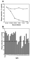
Monoclonal antibodies neutralize EEV. BSC-1 cell monolayers were infected with EEVs that were previously incubated with the indicated antibody. After 36 h of incubation at 37°C, the cells were fixed and stained with crystal violet. Data are expressed as the percentage of plaque reduction relative to the control with no IgG. (A) EEV neutralization using B5R(275t) mouse serum (black squares). An anti-A33R mouse serum was used as a negative control (open squares). (B) EEV reduction by anti-B5R(275t) MAbs at 25 μg/ml. An anti-myc monoclonal antibody was used as a negative control. The numbers on the x axis refer to the number of each VMC. The dashed line represents 60% of the control or 40% inhibition, and this value was used as a cutoff. Each value represents the mean of the percentage plaque number of triplicate measurements from two independent experiments.
FIG. 4.
FIG. 4.
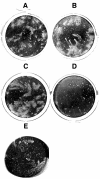
Inhibition of comet formation by anti-B5R(275t) MAbs. BSC-1 cell monolayers were infected with vaccinia virus strain IHDJ. Following adsorption, a liquid overlay containing 100 μg/ml of antibody was added. After 36 h of incubation at 37°C, the cultures were fixed and stained with crystal violet. Five representative results are shown: no antibody (A), anti-myc antibody used as a negative control (B), VMC-11 as an example of an anti-B5R(275t) antibody with no effect on comet formation (C), VMC-20 as an example of an anti-B5R(275t) antibody that inhibited comet formation (D), and rat MAb 19C2 at 20 μg/ml (E).
FIG. 5.
FIG. 5.
Overlapping peptides spanning the ectodomain of B5R. The amino acid sequence of B5R WR (amino acids 1 to 317) is indicated as a single letter code. The amino acids comprised by each peptide are represented as a black bar under the B5R sequence, and the peptide number is indicated alongside. Peptides are 20 amino acids in length and overlap the contiguous peptide by 10 amino acids, with the exception of peptide 28.
FIG. 6.
FIG. 6.
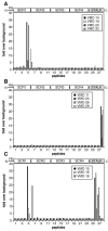
Peptide mapping of anti-B5R(275t) MAbs by ELISA. For each peptide, 10 pmol was immobilized on a 96-well streptavidin-coated plate and incubated with purified IgG (20 μg/ml). Bound IgG was detected with anti-mouse Ig-HRP and ABTS substrate. Absorbance was read at 405 nm. The numbers on the x axis refer to the number of each peptide. (A) Representative chart of MAbs binding to the area SCR1-SCR2. (B) Representative chart of MAbs binding to the stalk region. (C) MAbs binding to both the SCR1-SCR2 region and the stalk region.
FIG. 7.
FIG. 7.
Peptide mapping of anti-B5R(275t) MAbs. Recombinant B5R(275t) is illustrated at the top. The numbers above the diagram refer to the amino acid residues at the beginning or end of the feature depicted within the protein. Peptides (columns 1 to 28) spanning the extracellular domain of B5R were bound to streptavidin plates and probed by ELISA with each of the anti-B5R(275t) MAbs at 20 μg/ml (indicated in the second row). The failure of group 2D MAbs to recognize any peptide could be explained by failure of some biotinylated peptides to bind to the streptavidin on the ELISA plate, although this is highly unlikely. Alternatively, this failure could be due to their epitopes not being truly linear. A representative set of results is shown. Values within the box represent the change (n-fold) over background of each binding. Blank squares indicate values under 3-fold; gray indicates values between 3- and 10-fold; and black indicates values that were greater than 10-fold.
FIG. 8.
FIG. 8.
Analysis of VMC-18 biding to peptides. (A) Binding of VMC-18 to B5R(275t) after incubation with peptide 6 (left) or peptide 1 (right, used as a control). VMC-18 was incubated for 30 min at RT with 10 pmol (lane 2), 100 pmol (lane 3), 1 nmol (lane 4), or 10 nmol (lane 5) of peptide or with no peptide (lane 1). Purified baculovirus-expressed B5R(275t) was electrophoresed under denaturing conditions, transferred to nitrocellulose, and probed with the VMC-18-peptide mix. The sizes of molecular mass markers are shown in kilodaltons. (B) VMC-18 binding to peptide 6 (1), peptide 26 (2), or peptide 28 (3) after elution from peptide 6. Streptavidin-coated plates were coated with 10 pmol of peptide 6 and incubated with 20 μg/ml of VMC-18 for 1 h at RT. The wells were washed, and the bound VMC-18 was eluted with 0.1 M acid glycine, pH 2.6. After neutralization with a mixture of sodium carbonate and sodium bicarbonate, the eluate was added to wells coated with peptide 6, peptide 26, or peptide 28 (used as a control) for 1 h at RT. Bound IgG was detected with goat anti-mouse IgG coupled with HRP.
FIG. 9.
FIG. 9.
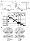
MAb blocking measured on a biosensor. (A) Example of a sensorgram obtained from two MAbs directed against independent epitopes. First, B5R(275t) (240 RU) was captured on the chip via an immobilized anti-His MAb (step 1), and then a primary antibody was allowed to bind to the captured B5R(275t) for 3 min (step 2). The secondary antibody (or test antibody) was then injected, and its association was monitored for another 3 min (step 3) (each MAb was injected at 20 μg/ml). Using this capture method, B5R(275t) is oriented “upside down” compared to the native type I protein. This may explain why 15 out of 26 MAbs (including neutralizing MAbs [Table 1]) failed to bind to B5R(275t) on the biosensor chip. In this example, the primary antibody was VMC-19 and the test antibody was VMC-23. (B) An overlaid sensorgram comprising only step 3 of the binding of test MAb VMC-20 to B5R(275t) after blocking with various primary antibodies is shown. The primary antibodies are indicated to the right. The sensorgrams are aligned at the time of injection of VMC-20. The binding of VMC-20 to the captured B5R(275t) in the absence of a primary antibody is labeled “control” and corresponds to 100% binding (0% blocking). Blocking of VMC-20 by itself (self) resulted in residual binding, which was considered to represent background. (C) The table indicates the percentage of blocking when each MAb (on the left) is injected after injection of the primary antibody (at the top). The MAbs are arranged in functional groups. The formula used to calculate the percentage of blocking is 100 − [(RUIgn − RUself) × 100/(RUcontrol − RUself)], where RUcontrol represents the binding in the absence of primary MAb, RUself represents residual binding after self-blocking, and RUIgn represents binding in the presence of primary MAb. This formula considers the RUcontrol to represent 100% binding and RUself to represent background normalized to zero. Black rectangles indicate high blocking (50 to 100%), gray rectangles indicate low blocking (40 to 50%), and white rectangles indicate no blocking. Reciprocal blocking was done only for competing pairs. (D) Diagram representation of blocking interactions between antibodies. Blocking is represented by a black line. The number to the top right of each antibody corresponds to the residues recognized by each antibody based on peptide mapping. Note that the blocking between the rat antibody and VMC-29 is not reciprocal, i.e., VMC-29 blocks the binding of the rat antibody, but the rat antibody does not block VMC-29 binding.
FIG. 10.
FIG. 10.
Models of the B5R protein. (A) B5R is divided into the SCR, stalk, transmembrane (TM), and cytoplasmic tail (CT) domains. It contains 317 amino acids, the first 19 amino acids being cleaved as the signal peptide (14). (B and C) Illustration of the potential B5R protein structure if the N- and C-terminal interaction were intramolecular (B) or intermolecular (C). Black lollipops represent putative N-linked carbohydrates.

References

    1. Appleyard, G., A. J. Hapel, and E. A. Boulter. 1971. An antigenic difference between intracellular and extracellular rabbitpox virus. J. Gen. Virol. 13:9-17. - PubMed
    1. Aslam, M., and S. J. Perkins. 2001. Folded-back solution structure of monomeric factor H of human complement by synchrotron X-ray and neutron scattering, analytical ultracentrifugation and constrained molecular modelling. J. Mol. Biol. 309:1117-1138. - PubMed
    1. Bell, E., M. Shamim, J. C. Whitbeck, G. Sfyroera, J. D. Lambris, and S. N. Isaacs. 2004. Antibodies against the extracellular enveloped virus B5R protein are mainly responsible for the EEV neutralizing capacity of vaccinia immune globulin. Virology 325:425-431. - PubMed
    1. Boulter, E. A., and G. Appleyard. 1973. Differences between extracellular and intracellular forms of poxvirus and their implications. Prog. Med. Virol. 16:86-108. - PubMed
    1. Bouma, B., P. G. de Groot, J. M. van den Elsen, R. B. Ravelli, A. Schouten, M. J. Simmelink, R. H. Derksen, J. Kroon, and P. Gros. 1999. Adhesion mechanism of human beta(2)-glycoprotein I to phospholipids based on its crystal structure. EMBO J. 18:5166-5174. - PMC - PubMed

Publication types

MeSH terms

LinkOut - more resources